Explore
 Validate
 Learn
Learn Immunocytochemistry
Antibody data
- Antibody Data
- Antigen structure
- References [3]
- Comments [0]
- Validations
- Immunocytochemistry [1]
- Immunohistochemistry [1]
Submit
Validation data
Reference
Comment
Report error
- Product number
- HPA013392 - Provider product page

- Provider
- Atlas Antibodies
- Proper citation
- Atlas Antibodies Cat#HPA013392, RRID:AB_1849468
- Product name
- Anti-GAP43
- Antibody type
- Polyclonal
- Description
- Polyclonal Antibody against Human GAP43, Gene description: growth associated protein 43, Alternative Gene Names: B-50, PP46, Validated applications: ICC, IHC, Uniprot ID: P17677, Storage: Store at +4°C for short term storage. Long time storage is recommended at -20°C.
- Reactivity
- Human
- Host
- Rabbit
- Conjugate
- Unconjugated
- Isotype
- IgG
- Vial size
- 100 µl
- Concentration
- 0.3 mg/ml
- Storage
- Store at +4°C for short term storage. Long time storage is recommended at -20°C.
- Handling
- The antibody solution should be gently mixed before use.
Submitted references Effect of transplantation of olfactory ensheathing cell conditioned medium induced bone marrow stromal cells on rats with spinal cord injury
CSF profiling of the human brain enriched proteome reveals associations of neuromodulin and neurogranin to Alzheimer's disease
Antibody‐based profiling of cerebrospinal fluid within multiple sclerosis
Feng L, Gan H, Zhao W, Liu Y
Molecular Medicine Reports 2017;16(2):1661-1668
Molecular Medicine Reports 2017;16(2):1661-1668
CSF profiling of the human brain enriched proteome reveals associations of neuromodulin and neurogranin to Alzheimer's disease
Remnestål J, Just D, Mitsios N, Fredolini C, Mulder J, Schwenk J, Uhlén M, Kultima K, Ingelsson M, Kilander L, Lannfelt L, Svenningsson P, Nellgård B, Zetterberg H, Blennow K, Nilsson P, Häggmark‐Månberg A
PROTEOMICS – Clinical Applications 2016;10(12):1242-1253
PROTEOMICS – Clinical Applications 2016;10(12):1242-1253
Antibody‐based profiling of cerebrospinal fluid within multiple sclerosis
Häggmark A, Byström S, Ayoglu B, Qundos U, Uhlén M, Khademi M, Olsson T, Schwenk J, Nilsson P
PROTEOMICS 2013;13(15):2256-2267
PROTEOMICS 2013;13(15):2256-2267
No comments: Submit comment
Supportive validation
- Submitted by
- Atlas Antibodies (provider)
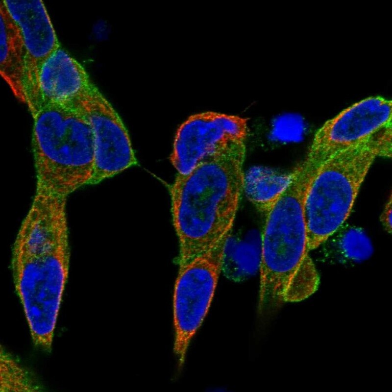
- Main image

- Experimental details
- Immunofluorescent staining of human cell line SH-SY5Y shows localization to plasma membrane.
- Sample type
- Human
Supportive validation
- Submitted by
- Atlas Antibodies (provider)
- Enhanced method
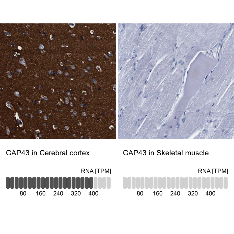
- Orthogonal validation
- Main image

- Experimental details
- Immunohistochemistry analysis in human cerebral cortex and skeletal muscle tissues using HPA013392 antibody. Corresponding GAP43 RNA-seq data are presented for the same tissues.
- Sample type
- Human
- Protocol
- Protocol