 Explore
 Validate
 Learn
Learn Western blot
Antibody data
- Antibody Data
- Antigen structure
- References [1]
- Comments [0]
- Validations
- Western blot [2]
- Immunohistochemistry [1]
Submit
Validation data
Reference
Comment
Report error
- Product number
- AF3580 - Provider product page

- Provider
- Novus Biologicals
- Product name
- Goat Polyclonal FKBP38 Antibody
- Antibody type
- Polyclonal
- Description
- Immunogen affinity purified. Detects human, mouse, and rat FKBP38 in Western blots. In Western blots, less than 1% cross-reactivity with recombinant human FKBP-12, -12.6, -13, -25, -51, or -52 is observed.
- Reactivity
- Human, Mouse, Rat
- Host
- Goat
- Conjugate
- Unconjugated
- Isotype
- IgG
- Vial size
- 100 ug
- Concentration
- LYOPH
- Storage
- Use a manual defrost freezer and avoid repeated freeze-thaw cycles. 12 months from date of receipt, -20 to -70 degreesC as supplied. 1 month, 2 to 8 degreesC under sterile conditions after reconstitution. 6 months, -20 to -70 degreesC under sterile conditions after reconstitution.
Submitted references Hepatitis C virus NS5A activates the mammalian target of rapamycin (mTOR) pathway, contributing to cell survival by disrupting the interaction between FK506-binding protein 38 (FKBP38) and mTOR.
Peng L, Liang D, Tong W, Li J, Yuan Z
The Journal of biological chemistry 2010 Jul 2;285(27):20870-81
The Journal of biological chemistry 2010 Jul 2;285(27):20870-81
No comments: Submit comment
Supportive validation
- Submitted by
- Novus Biologicals (provider)
- Main image
- Experimental details
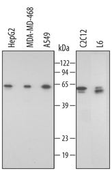
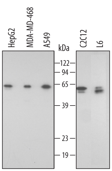
- Detection of Human/Mouse/Rat FKBP38 by Western Blot. Western blot shows lysates of HepG2 human hepatocellular carcinoma cell line, MDA-MB-468 human breast cancer cell line, A549 human lung carcinoma cell line, C2C12 mouse myoblast cell line, and L6 rat myoblast cell line. PVDF membrane was probed with 0.5 µg/mL of Goat Anti-Human/Mouse/Rat FKBP38 Antigen Affinity-purified Polyclonal Antibody (Catalog # AF3580) followed by HRP-conjugated Anti-Goat IgG Secondary Antibody (Catalog # HAF109). A specific band was detected for FKBP38 at approximately 60 - 64 kDa (as indicated). This experiment was conducted under reducing conditions and using Immunoblot Buffer Group 2.
- Submitted by
- Novus Biologicals (provider)
- Main image
- Experimental details
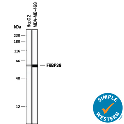
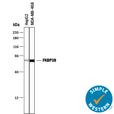
- Detection of Human FKBP38 by Simple WesternTM. Simple Western lane view shows lysates of HepG2 human hepatocellular carcinoma cell line and MDA-MB-468 human breast cancer cell line, loaded at 0.2 mg/mL. A specific band was detected for FKBP38 at approximately 58 kDa (as indicated) using 5 µg/mL of Goat Anti-Human/Mouse/Rat FKBP38 Antigen Affinity-purified Polyclonal Antibody (Catalog # AF3580) followed by 1:50 dilution of HRP-conjugated Anti-Goat IgG Secondary Antibody (Catalog # HAF109). This experiment was conducted under reducing conditions and using the 12-230 kDa separation system.
Supportive validation
- Submitted by
- Novus Biologicals (provider)
- Main image
- Experimental details
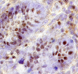
- FKBP38 in Human Intestine. FKBP38 was detected in immersion fixed paraffin-embedded sections of human intestine using 15 µg/mL Goat Anti-Human/Mouse/Rat FKBP38 Antigen Affinity-purified Polyclonal Antibody (Catalog # AF3580) overnight at 4 °C. Tissue was stained with the Anti-Goat HRP-DAB Cell & Tissue Staining Kit (brown; Catalog # CTS008) and counterstained with hematoxylin (blue). Specific labeling was localized to the nucleus of epithelial cells in intestinal glands. View our protocol for Chromogenic IHC Staining of Paraffin-embedded Tissue Sections. This application has not been tested in rat or mouse tissue.
