Explore
 Validate
 Learn
Learn Western blot
Antibody data
- Antibody Data
- Antigen structure
- References [0]
- Comments [0]
- Validations
- Western blot [1]
- Immunocytochemistry [1]
- Immunohistochemistry [1]
Submit
Validation data
Reference
Comment
Report error
- Product number
- MAB5745R-100 - Provider product page

- Provider
- R&D Systems
- Product name
- Human gamma-Synuclein Antibody
- Antibody type
- Monoclonal
- Description
- Protein A or G purified from cell culture supernatant. Detects human gamma-Synuclein in direct ELISAs and Western blots.
- Reactivity
- Human
- Host
- Mouse
- Conjugate
- Unconjugated
- Antigen sequence
Q6FHG5- Isotype
- IgG
- Antibody clone number
- 514304R
- Vial size
- 100 ug
- Storage
- Use a manual defrost freezer and avoid repeated freeze-thaw cycles. 12 months from date of receipt, -20 to -70 °C as supplied. 1 month, 2 to 8 °C under sterile conditions after reconstitution. 6 months, -20 to -70 °C under sterile conditions after reconstitution.
No comments: Submit comment
Supportive validation
- Submitted by
- R&D Systems (provider)
- Main image
- Experimental details
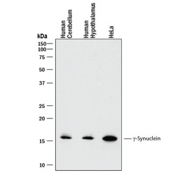
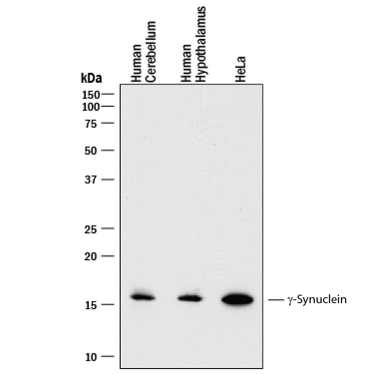
- Detection of Human gamma-Synuclein by Western Blot. Western blot shows lysates of human brain (cerebellum) tissue, human brain (hypothalamus) tissue, and HeLa human cervical epithelial carcinoma cell line. PVDF membrane was probed with 0.5 µg/mL of Recombinant Mouse Anti-Human gamma-Synuclein Monoclonal Antibody (Catalog # MAB5745R) followed by HRP-conjugated Anti-Mouse IgG Secondary Antibody (Catalog # HAF018). A specific band was detected for gamma-Synuclein at approximately 16 kDa (as indicated). This experiment was conducted under reducing conditions and using Immunoblot Buffer Group 1.
Supportive validation
- Submitted by
- R&D Systems (provider)
- Main image
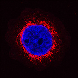
- Experimental details
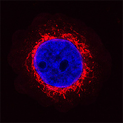
- gamma-Synuclein in MBA-MB-468 Human Cell Line. gamma-Synuclein was detected in immersion fixed MBA-MB-468 human breast cancer cell line using Recombinant Mouse Anti-Human gamma-Synuclein Monoclonal Antibody (Catalog # MAB5745R) at 1.7 µg/mL for 3 hours at room temperature. Cells were stained using the NorthernLights™ 557-conjugated Anti-Mouse IgG Secondary Antibody (red; Catalog # NL007) and counterstained with DAPI (blue). Specific staining was localized to cytoplasm. View our protocol for Fluorescent ICC Staining of Non-adherent Cells.
Supportive validation
- Submitted by
- R&D Systems (provider)
- Main image
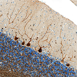
- Experimental details
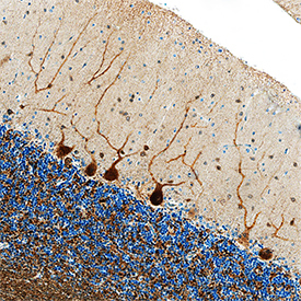
- gamma-Synuclein in Human Brain. gamma-Synuclein was detected in immersion fixed paraffin-embedded sections of human brain (cerebellum) using Recombinant Mouse Anti-Human gamma-Synuclein Monoclonal Antibody (Catalog # MAB5745R) at 1 µg/mL overnight at 4 °C. Tissue was stained using the Anti-Mouse HRP-DAB Cell & Tissue Staining Kit (brown; Catalog # CTS002) and counterstained with hematoxylin (blue). Specific staining was localized to Purkinje neurons. View our protocol for Chromogenic IHC Staining of Paraffin-embedded Tissue Sections.