Explore
 Validate
 Learn
Learn Western blot
 Flow cytometry
Antibody data
- Antibody Data
- Antigen structure
- References [0]
- Comments [0]
- Validations
- Flow cytometry [2]
Submit
Validation data
Reference
Comment
Report error
- Product number
- PA5-118397 - Provider product page

- Provider
- Invitrogen Antibodies
- Product name
- CPEB1 Polyclonal Antibody
- Antibody type
- Polyclonal
- Antigen
- Synthetic peptide
- Reactivity
- Human, Mouse, Rat
- Host
- Rabbit
- Isotype
- IgG
- Vial size
- 100 µL
- Concentration
- 1 mg/mL
- Storage
- Store at 4°C short term. For long term storage, store at -20°C, avoiding freeze/thaw cycles.
No comments: Submit comment
Supportive validation
- Submitted by
- Invitrogen Antibodies (provider)
- Main image
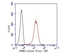
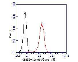
- Experimental details
- Flow Cytometry analysis of CPEB1 in K562 cells. The cells were fixed, permeabilized and stained with CPEB1 polyclonal Antibody (Product # PA5-118397) at a dilution of 1:50 (red). After incubation of the primary antibody at room temperature for an hour, the cells were stained with a Alexa Fluor 488-conjugated Goat anti-Rabbit IgG Secondary antibody at 1:1,000 dilution for 30 minutes. Unlabelled sample was used as a control (cells without incubation with primary antibody; black).
- Submitted by
- Invitrogen Antibodies (provider)
- Main image
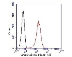
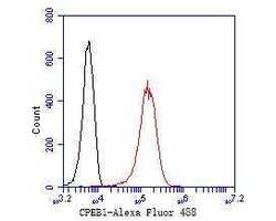
- Experimental details
- Flow Cytometry analysis of CPEB1 in K562 cells. The cells were fixed, permeabilized and stained with CPEB1 polyclonal Antibody (Product # PA5-118397) at a dilution of 1:50 (red). After incubation of the primary antibody at room temperature for an hour, the cells were stained with a Alexa Fluor 488-conjugated Goat anti-Rabbit IgG Secondary antibody at 1:1,000 dilution for 30 minutes. Unlabelled sample was used as a control (cells without incubation with primary antibody; black).