Explore
 Validate
 Learn
Learn Western blot
Antibody data
- Antibody Data
- Antigen structure
- References [3]
- Comments [0]
- Validations
- Western blot [1]
- Immunohistochemistry [1]
- Blocking/Neutralizing [1]
Submit
Validation data
Reference
Comment
Report error
- Product number
- AF1560 - Provider product page

- Provider
- Novus Biologicals
- Product name
- Goat Polyclonal PDGF-C Antibody
- Antibody type
- Polyclonal
- Description
- Immunogen affinity purified. Detects human PDGF-C in direct ELISAs and Western blots. In Western blots, approximately 15% cross-reactivity with recombinant mouse PDGF-C is observed, and less than 1% cross-reactivity with human PDGF, recombinant human (rh) PDGF-AA, rhPDGF-BB, rhPDGF-AB, and rhPDGF-D is observed.
- Reactivity
- Human
- Host
- Goat
- Isotype
- IgG
- Vial size
- 100 ug
- Concentration
- LYOPH
- Storage
- Use a manual defrost freezer and avoid repeated freeze-thaw cycles. 12 months from date of receipt, -20 to -70 degreesC as supplied. 1 month, 2 to 8 degreesC under sterile conditions after reconstitution. 6 months, -20 to -70 degreesC under sterile conditions after reconstitution.
Submitted references PDGF-CC underlies resistance to VEGF-A inhibition and combinatorial targeting of both suppresses pathological angiogenesis more efficiently.
Platelet-derived growth factor C and calpain-3 are modulators of human melanoma cell invasiveness.
Effects of PDGF-C and PDGF-D on monocyte migration and MMP-2 and MMP-9 expression.
Zheng L, Zhao C, Du Y, Lin X, Jiang Y, Lee C, Tian G, Mi J, Li X, Chen Q, Ye Z, Huang L, Wang S, Ren X, Xing L, Chen W, Huang D, Gao Z, Zhang S, Lu W, Tang Z, Wang B, Ju R, Li X
Oncotarget 2016 Nov 22;7(47):77902-77915
Oncotarget 2016 Nov 22;7(47):77902-77915
Platelet-derived growth factor C and calpain-3 are modulators of human melanoma cell invasiveness.
Ruffini F, Tentori L, Dorio AS, Arcelli D, D'Amati G, D'Atri S, Graziani G, Lacal PM
Oncology reports 2013 Dec;30(6):2887-96
Oncology reports 2013 Dec;30(6):2887-96
Effects of PDGF-C and PDGF-D on monocyte migration and MMP-2 and MMP-9 expression.
Wågsäter D, Zhu C, Björck HM, Eriksson P
Atherosclerosis 2009 Feb;202(2):415-23
Atherosclerosis 2009 Feb;202(2):415-23
No comments: Submit comment
Supportive validation
- Submitted by
- Novus Biologicals (provider)
- Main image
- Experimental details
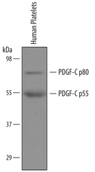
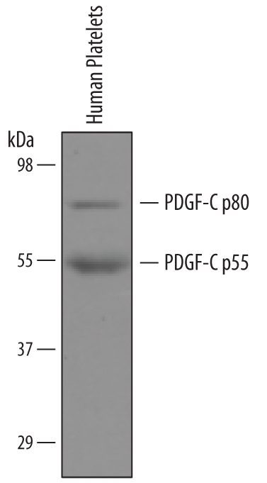
- Detection of Human PDGF-C by Western Blot. Western blot shows lysates of human platelets. PVDF membrane was probed with 1 µg/mL of Goat Anti-Human PDGF-C Antigen Affinity-purified Polyclonal Antibody (Catalog # AF1560) followed by HRP-conjugated Anti-Goat IgG Secondary Antibody (Catalog # HAF109). Specific bands were detected for PDGF-C p80 at approximately 80 kDa and PDGF-C p55 at approximately 55 kDa (as indicated). This experiment was conducted under reducing conditions and using Immunoblot Buffer Group 8.
Supportive validation
- Submitted by
- Novus Biologicals (provider)
- Main image
- Experimental details
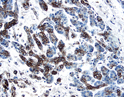
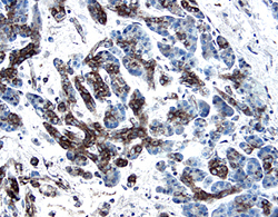
- PDGF-C in Human Pancreatic Cancer Tissue. PDGF-C was detected in immersion fixed paraffin-embedded sections of human pancreatic cancer tissue using 1.7 µg/mL Goat Anti-Human PDGF-C Antigen Affinity-purified Polyclonal Antibody (Catalog # AF1560) overnight at 4 °C. Tissue was stained with the Anti-Goat HRP-DAB Cell & Tissue Staining Kit (brown; Catalog # CTS008) and counterstained with hematoxylin (blue). View our protocol for Chromogenic IHC Staining of Paraffin-embedded Tissue Sections.
Supportive validation
- Submitted by
- Novus Biologicals (provider)
- Main image
- Experimental details
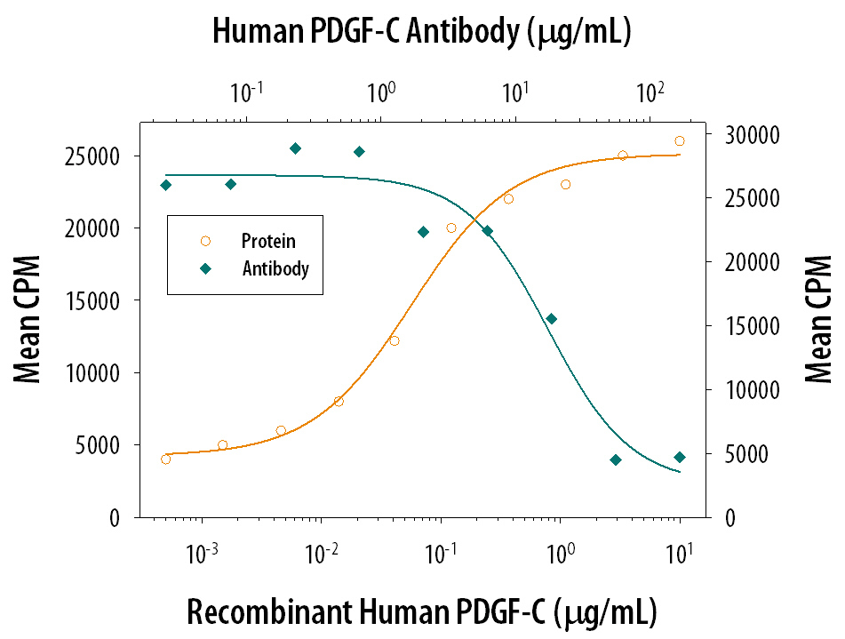
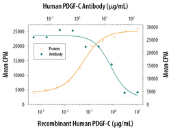
- Cell Proliferation Induced by PDGF-CC and Neutralization by Human PDGF-C Antibody. Recombinant Human PDGF-CC (Catalog # 1687-CC) stimulates proliferation in the NR6R-3T3 mouse fibroblast cell line in a dose-dependent manner (orange line). Proliferation elicited by Recombinant Human PDGF-CC (0.8 µg/mL) is neutralized (green line) by increasing concentrations of Goat Anti-Human PDGF-C Antigen Affinity-purified Polyclonal Antibody (Catalog # AF1560). The ND50 is typically 6-24 µg/mL.