 Explore
 Validate
 Learn
Learn Western blot
 Immunohistochemistry
Antibody data
- Antibody Data
- Antigen structure
- References [0]
- Comments [0]
- Validations
- Immunohistochemistry [1]
- Other assay [2]
Submit
Validation data
Reference
Comment
Report error
- Product number
- PA5-47156 - Provider product page

- Provider
- Invitrogen Antibodies
- Product name
- PDGF-C Polyclonal Antibody
- Antibody type
- Polyclonal
- Antigen
- Recombinant full-length protein
- Description
- In Western blots, approximately 15% cross-reactivity with recombinant mouse PDGF-C is observed, and less than 1% cross-reactivity with human PDGF, recombinant human (rh) PDGF-AA, rhPDGF-BB, rhPDGF-AB, and rhPDGF-D is observed. Reconstitute at 0.2 mg/mL in sterile PBS. Endoxin level is
- Reactivity
- Human
- Host
- Goat
- Isotype
- IgG
- Vial size
- 100 μg
- Concentration
- 0.2 mg/mL
- Storage
- -20°C, Avoid Freeze/Thaw Cycles
No comments: Submit comment
Supportive validation
- Submitted by
- Invitrogen Antibodies (provider)
- Main image

- Experimental details
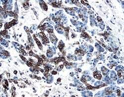
- Immunohistochemical analysis of PDGF-C in immersion fixed paraffin-embedded sections of human pancreatic cancer tissue. Samples were incubated in PDGF-C polyclonal antibody (Product # PA5-47156) using a dilution of 1.7 µg/mL overnight at 4 °C. Tissue was stained with the Anti-Goat HRP-DAB Cell & Tissue Staining Kit (brown) and counterstained with hematoxylin (blue).
Supportive validation
- Submitted by
- Invitrogen Antibodies (provider)
- Main image
- Experimental details
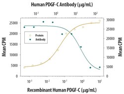
- Neutralization of PDGF-C in NR6R‚3T3 mouse fibroblast cell line. Samples were incubated in PDGF-C polyclonal antibody (Product # PA5-47156). Recombinant Human PDGF‚CC stimulates proliferation in the NR6R‚3T3 mouse fibroblast cell line in a dose-dependent manner (orange line). Proliferation elicited by Recombinant Human PDGF‚CC (0.8 µg/mL) is neutralized (green line) by increasing concentrations of Goat Anti-Human PDGF-C Antigen Affinity-purified Polyclonal Antibody. The ND50 is typically 6-24 µg/mL.
- Submitted by
- Invitrogen Antibodies (provider)
- Main image

- Experimental details
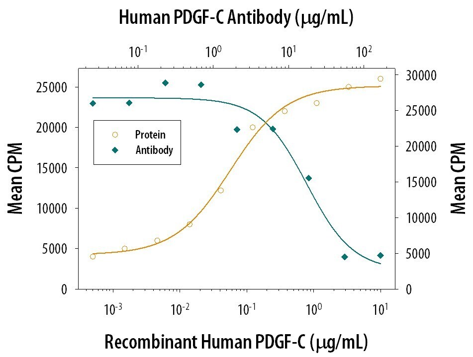
- Neutralization of PDGF-C in NR6R‚3T3 mouse fibroblast cell line. Samples were incubated in PDGF-C polyclonal antibody (Product # PA5-47156). Recombinant Human PDGF‚CC stimulates proliferation in the NR6R‚3T3 mouse fibroblast cell line in a dose-dependent manner (orange line). Proliferation elicited by Recombinant Human PDGF‚CC (0.8 µg/mL) is neutralized (green line) by increasing concentrations of Goat Anti-Human PDGF-C Antigen Affinity-purified Polyclonal Antibody. The ND50 is typically 6-24 µg/mL.
