 Explore
 Validate
 Learn
Learn Western blot
 Immunocytochemistry
Antibody data
- Antibody Data
- Antigen structure
- References [12]
- Comments [0]
- Validations
- Western blot [10]
- Immunocytochemistry [8]
- Immunoprecipitation [1]
- Immunohistochemistry [3]
Submit
Validation data
Reference
Comment
Report error
- Product number
- GTX110624 - Provider product page

- Provider
- GeneTex
- Proper citation
- GeneTex Cat#GTX110624, RRID:AB_1950045
- Product name
- Citrate synthetase antibody [N2C3]
- Antibody type
- Polyclonal
- Reactivity
- Human, Mouse, Rat, Chicken/Avian, Simian
- Host
- Rabbit
Submitted references Mild inborn errors of metabolism in commonly used inbred mouse strains.
Effects of long-term exposures to low iron and branched-chain amino acid containing diets on aging skeletal muscle of Fisher 344 × Brown Norway rats.
Myostatin deficiency is associated with lipidomic abnormalities in skeletal muscles.
Mitochondrial retrograde signaling connects respiratory capacity to thermogenic gene expression.
Mitochondrial quality-control dysregulation in conditional HO-1(-/-) mice.
Biallelic Mutations in MRPS34 Lead to Instability of the Small Mitoribosomal Subunit and Leigh Syndrome.
Ataxin-1 regulates the cerebellar bioenergetics proteome through the GSK3β-mTOR pathway which is altered in Spinocerebellar ataxia type 1 (SCA1).
Effects of inhaled CO administration on acute lung injury in baboons with pneumococcal pneumonia.
Redox regulation of mitophagy in the lung during murine Staphylococcus aureus sepsis.
Changes in peak fat oxidation in response to different doses of endurance training.
SIRT3, a mitochondrial NAD⁺-dependent deacetylase, is involved in the regulation of myoblast differentiation.
The birth of quail chicks after intracytoplasmic sperm injection.
Leandro J, Violante S, Argmann CA, Hagen J, Dodatko T, Bender A, Zhang W, Williams EG, Bachmann AM, Auwerx J, Yu C, Houten SM
Molecular genetics and metabolism 2019 Apr;126(4):388-396
Molecular genetics and metabolism 2019 Apr;126(4):388-396
Effects of long-term exposures to low iron and branched-chain amino acid containing diets on aging skeletal muscle of Fisher 344 × Brown Norway rats.
Kim Y, Men SS, Liang C, Receno CN, Brutsaert TD, Korol DL, Heffernan KS, DeRuisseau KC
Applied physiology, nutrition, and metabolism = Physiologie appliquee, nutrition et metabolisme 2018 Feb;43(2):165-173
Applied physiology, nutrition, and metabolism = Physiologie appliquee, nutrition et metabolisme 2018 Feb;43(2):165-173
Myostatin deficiency is associated with lipidomic abnormalities in skeletal muscles.
Baati N, Feillet-Coudray C, Fouret G, Vernus B, Goustard B, Coudray C, Lecomte J, Blanquet V, Magnol L, Bonnieu A, Koechlin-Ramonatxo C
Biochimica et biophysica acta. Molecular and cell biology of lipids 2017 Oct;1862(10 Pt A):1044-1055
Biochimica et biophysica acta. Molecular and cell biology of lipids 2017 Oct;1862(10 Pt A):1044-1055
Mitochondrial retrograde signaling connects respiratory capacity to thermogenic gene expression.
Nam M, Akie TE, Sanosaka M, Craige SM, Kant S, Keaney JF Jr, Cooper MP
Scientific reports 2017 May 17;7(1):2013
Scientific reports 2017 May 17;7(1):2013
Mitochondrial quality-control dysregulation in conditional HO-1(-/-) mice.
Suliman HB, Keenan JE, Piantadosi CA
JCI insight 2017 Feb 9;2(3):e89676
JCI insight 2017 Feb 9;2(3):e89676
Biallelic Mutations in MRPS34 Lead to Instability of the Small Mitoribosomal Subunit and Leigh Syndrome.
Lake NJ, Webb BD, Stroud DA, Richman TR, Ruzzenente B, Compton AG, Mountford HS, Pulman J, Zangarelli C, Rio M, Boddaert N, Assouline Z, Sherpa MD, Schadt EE, Houten SM, Byrnes J, McCormick EM, Zolkipli-Cunningham Z, Haude K, Zhang Z, Retterer K, Bai R, Calvo SE, Mootha VK, Christodoulou J, Rötig A, Filipovska A, Cristian I, Falk MJ, Metodiev MD, Thorburn DR
American journal of human genetics 2017 Aug 3;101(2):239-254
American journal of human genetics 2017 Aug 3;101(2):239-254
Ataxin-1 regulates the cerebellar bioenergetics proteome through the GSK3β-mTOR pathway which is altered in Spinocerebellar ataxia type 1 (SCA1).
Sánchez I, Balagué E, Matilla-Dueñas A
Human molecular genetics 2016 Sep 15;25(18):4021-4040
Human molecular genetics 2016 Sep 15;25(18):4021-4040
Effects of inhaled CO administration on acute lung injury in baboons with pneumococcal pneumonia.
Fredenburgh LE, Kraft BD, Hess DR, Harris RS, Wolf MA, Suliman HB, Roggli VL, Davies JD, Winkler T, Stenzler A, Baron RM, Thompson BT, Choi AM, Welty-Wolf KE, Piantadosi CA
American journal of physiology. Lung cellular and molecular physiology 2015 Oct 15;309(8):L834-46
American journal of physiology. Lung cellular and molecular physiology 2015 Oct 15;309(8):L834-46
Redox regulation of mitophagy in the lung during murine Staphylococcus aureus sepsis.
Chang AL, Ulrich A, Suliman HB, Piantadosi CA
Free radical biology & medicine 2015 Jan;78:179-89
Free radical biology & medicine 2015 Jan;78:179-89
Changes in peak fat oxidation in response to different doses of endurance training.
Rosenkilde M, Reichkendler MH, Auerbach P, Bonne TC, Sjödin A, Ploug T, Stallknecht BM
Scandinavian journal of medicine & science in sports 2015 Feb;25(1):41-52
Scandinavian journal of medicine & science in sports 2015 Feb;25(1):41-52
SIRT3, a mitochondrial NAD⁺-dependent deacetylase, is involved in the regulation of myoblast differentiation.
Abdel Khalek W, Cortade F, Ollendorff V, Lapasset L, Tintignac L, Chabi B, Wrutniak-Cabello C
PloS one 2014;9(12):e114388
PloS one 2014;9(12):e114388
The birth of quail chicks after intracytoplasmic sperm injection.
Mizushima S, Hiyama G, Shiba K, Inaba K, Dohra H, Ono T, Shimada K, Sasanami T
Development (Cambridge, England) 2014 Oct;141(19):3799-806
Development (Cambridge, England) 2014 Oct;141(19):3799-806
No comments: Submit comment
Enhanced validation
Supportive validation
- Submitted by
- GeneTex (provider)
- Enhanced method
- Genetic validation
- Main image
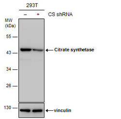
- Experimental details
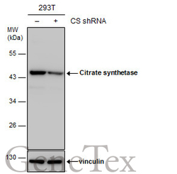
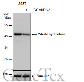
- Non-transfected (¡V) and transfected (+) 293T whole cell extracts (30 ?g) were separated by 10% SDS-PAGE, and the membrane was blotted with Citrate synthetase antibody [N2C3] (GTX110624) diluted at 1:1000. The HRP-conjugated anti-rabbit IgG antibody (GTX213110-01) was used to detect the primary antibody.
Supportive validation
- Submitted by
- GeneTex (provider)
- Main image
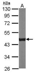
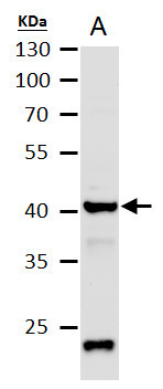
- Experimental details
- Sample (50 ug of whole cell lysate) A: Mouse brain 10% SDS PAGE GTX110624 diluted at 1:1000
- Validation comment
- WB

- Submitted by
- GeneTex (provider)
- Main image
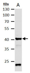
- Experimental details
- Citrate synthetase antibody [N2C3] detects Citrate synthetase protein by western blot analysis.A. 50 ?g mouse brain extract10 % SDS-PAGECitrate synthetase antibody [N2C3] (GTX110624) dilution: 1:1000
- Validation comment
- WB
- Submitted by
- GeneTex (provider)
- Main image
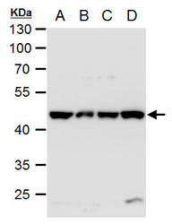
- Experimental details
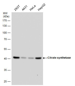
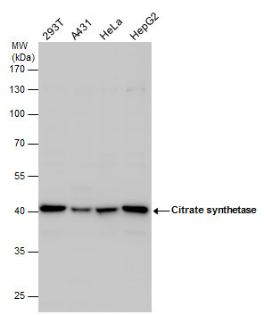
- Citrate synthetase antibody [N2C3] detects Citrate synthetase protein by western blot analysis.A. 30 ?g 293T whole cell extractB. 30 ?g A431 whole cell extractC. 30 ?g HeLa whole cell extractD. 30 ?g HepG2 whole cell extract10 % SDS-PAGECitrate synthetase antibody [N2C3] (GTX110624) dilution: 1:1000
- Validation comment
- WB

- Submitted by
- GeneTex (provider)
- Main image
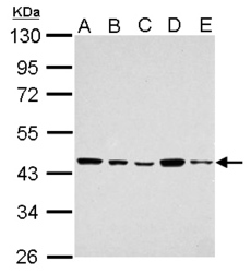
- Experimental details
- Citrate synthetase antibody [N2C3] detects Citrate synthetase protein by western blot analysis.A. 30 ?g 293T whole cell lysate/extractB. 30 ?g A431 whole cell lysate/extractC. 30 ?g HeLa whole cell lysate/extractD. 30 ?g HepG2 whole cell lysate/extractE. 30 ?g A375 whole cell lysate/extract10 % SDS-PAGECitrate synthetase antibody [N2C3] (GTX110624) dilution: 1:1000
- Validation comment
- WB
- Submitted by
- GeneTex (provider)
- Main image
- Experimental details
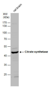
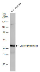
- Citrate synthetase antibody detects Citrate synthetase protein by western blot analysis. Rat tissue extracts (50 ?g) was separated by 10% SDS-PAGE, and the membrane was blotted with Citrate synthetase antibody (GTX110624) diluted by 1:1000. The HRP-conjugated anti-rabbit IgG antibody (GTX213110-01) was used to detect the primary antibody.
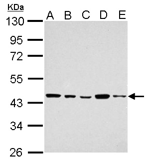
- Submitted by
- GeneTex (provider)
- Main image

- Experimental details
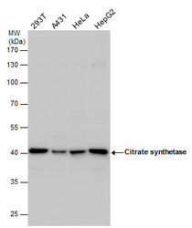
- Citrate synthetase antibody detects Citrate synthetase protein by western blot analysis. Various whole cell extracts (30 ?g) were separated by 10% SDS-PAGE, and the membrane was blotted with Citrate synthetase antibody (GTX110624) diluted by 1:1000. The HRP-conjugated anti-rabbit IgG antibody (GTX213110-01) was used to detect the primary antibody.
- Submitted by
- GeneTex (provider)
- Main image
- Experimental details
- Citrate synthetase antibody detects Citrate synthetase protein by Western blot analysis. Various whole cell extracts (30 £gg) were separated by 10% SDS-PAGE, and the membrane was blotted with Citrate synthetase antibody (GTX110624) diluted at a dilution of 1:1000.
- Submitted by
- GeneTex (provider)
- Main image
- Experimental details
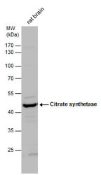
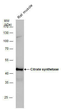
- Citrate synthetase antibody detects Citrate synthetase protein by Western blot analysis. Rat tissue extracts (50 £gg) was separated by 10 % SDS-PAGE, and the membrane was blotted with Citrate synthetase antibody (GTX110624) at a dilution of 1:1000.
- Submitted by
- GeneTex (provider)
- Main image
- Experimental details
- Non-transfected (¡V) and transfected (+) 293T whole cell extracts (30 ?g) were separated by 10% SDS-PAGE, and the membrane was blotted with Citrate synthetase antibody [N2C3] (GTX110624) diluted at 1:1000. The HRP-conjugated anti-rabbit IgG antibody (GTX213110-01) was used to detect the primary antibody.
Supportive validation
- Submitted by
- GeneTex (provider)
- Main image
- Experimental details
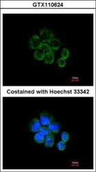
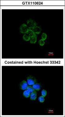
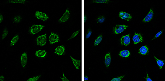
- Immunofluorescence analysis of methanol-fixed A431, using Citrate synthetase(GTX110624) antibody at 1:200 dilution.
- Submitted by
- GeneTex (provider)
- Main image
- Experimental details
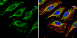
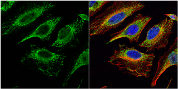
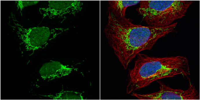
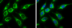
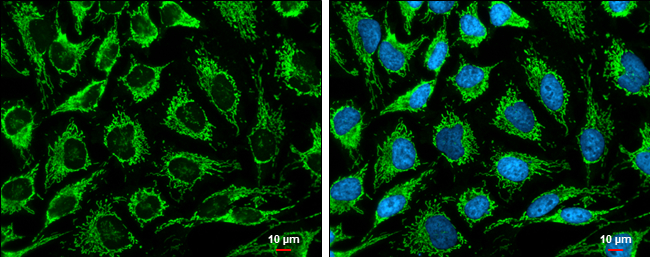
- Citrate synthetase antibody [N2C3] detects Citrate synthetase protein at mitochondria by immunofluorescent analysis.Sample: HeLa cells were fixed in 4% paraformaldehyde at RT for 15 min.Green: Citrate synthetase protein stained by Citrate synthetase antibody [N2C3] (GTX110624) diluted at 1:200.Red: alpha Tubulin, a cytoskeleton marker, stained by alpha Tubulin antibody [GT114] (GTX628802) diluted at 1:1000.Blue: Hoechst 33342 staining.
- Submitted by
- GeneTex (provider)
- Main image
- Experimental details
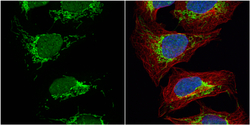
- Citrate synthetase antibody [N2C3] detects Citrate synthetase protein at mitochondria by immunofluorescent analysis.Sample: U2OS cells were fixed in 4% paraformaldehyde at RT for 15 min.Green: Citrate synthetase protein stained by Citrate synthetase antibody [N2C3] (GTX110624) diluted at 1:500.Red: alpha Tubulin, a cytoskeleton marker, stained by alpha Tubulin antibody [GT114] (GTX628802) diluted at 1:1000.Blue: Hoechst 33342 staining.
- Submitted by
- GeneTex (provider)
- Main image
- Experimental details
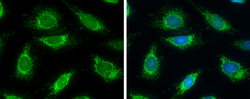
- Citrate synthetase antibody [N2C3] detects Citrate synthetase protein at mitochondria by immunofluorescent analysis.Sample: HeLa cells were fixed in 4% paraformaldehyde at RT for 15 min.Green: Citrate synthetase protein stained by Citrate synthetase antibody [N2C3] (GTX110624) diluted at 1:500.Blue: Hoechst 33342 staining.
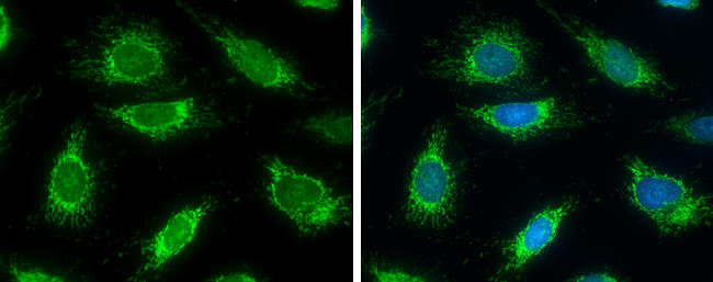
- Submitted by
- GeneTex (provider)
- Main image

- Experimental details
- Citrate synthetase antibody [N2C3] detects Citrate synthetase protein at mitochondria by immunofluorescent analysis.Sample: HeLa cells were fixed in ice-cold MeOH for 5 min.Green: Citrate synthetase protein stained by Citrate synthetase antibody [N2C3] (GTX110624) diluted at 1:500.Blue: Hoechst 33342 staining.
- Submitted by
- GeneTex (provider)
- Main image
- Experimental details
- Citrate synthetase antibody [N2C3] detects Citrate synthetase protein at mitochondria by immunofluorescent analysis.Sample: HeLa cells were fixed in 4% paraformaldehyde at RT for 15 min.Green: Citrate synthetase stained by Citrate synthetase antibody [N2C3] (GTX110624) diluted at 1:500.Blue: Hoechst 33342 staining.
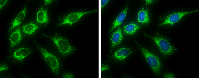
- Submitted by
- GeneTex (provider)
- Main image

- Experimental details
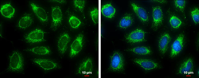
- Citrate synthetase antibody [N2C3] detects Citrate synthetase protein at mitochondria by immunofluorescent analysis.Sample: HeLa cells were fixed in 4% paraformaldehyde at RT for 15 min.Green: Citrate synthetase stained by Citrate synthetase antibody [N2C3] (GTX110624) diluted at 1:500.Blue: Hoechst 33342 staining.Scale bar= 10£gm.
- Submitted by
- GeneTex (provider)
- Main image

- Experimental details
- Citrate synthetase antibody [N2C3] detects Citrate synthetase protein at mitochondria by immunofluorescent analysis.Sample: HeLa cells were fixed in 4% paraformaldehyde at RT for 15 min.Green: Citrate synthetase stained by Citrate synthetase antibody [N2C3] (GTX110624) diluted at 1:500.Blue: Hoechst 33342 staining.Scale bar= 10£gm.
Supportive validation
- Submitted by
- GeneTex (provider)
- Main image
- Experimental details
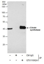
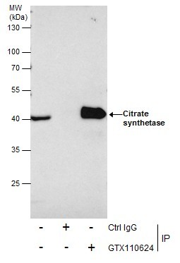
- Immunoprecipitation of Citrate synthetase protein from 293T whole cell extracts using 5 £gg of Citrate synthetase antibody [N2C3] (GTX110624).Western blot analysis was performed using Citrate synthetase antibody [N2C3] (GTX110624).EasyBlot anti-Rabbit IgG (GTX221666-01) was used as a secondary reagent.
Supportive validation
- Submitted by
- GeneTex (provider)
- Main image

- Experimental details
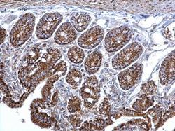
- Immunohistochemical analysis of paraffin-embedded NCI-N87 xenograft, using Citrate synthetase(GTX110624) antibody at 1:500 dilution.
- Submitted by
- GeneTex (provider)
- Main image
- Experimental details
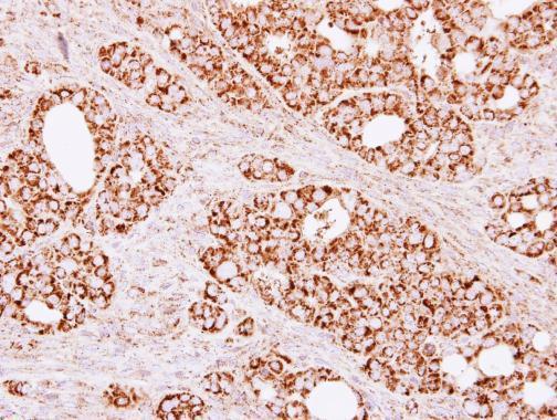
- Citrate synthetase antibody [N2C3] detects Citrate synthetase protein at mitochondria on mouse intestine by immunohistochemical analysis. Sample: Paraffin-embedded mouse intestine. Citrate synthetase antibody [N2C3] (GTX110624) dilution: 1:500.

- Submitted by
- GeneTex (provider)
- Main image
- Experimental details
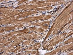
- Citrate synthetase antibody [N2C3] detects Citrate synthetase protein at cytoplasm in mouse muscle by immunohistochemical analysis. Sample: Paraffin-embedded mouse muscle. Citrate synthetase antibody [N2C3] (GTX110624) diluted at 1:500.
