 Explore
 Validate
 Learn
Learn Western blot
Antibody data
- Antibody Data
- Antigen structure
- References [0]
- Comments [0]
- Validations
- Western blot [1]
- Immunocytochemistry [1]
- Immunohistochemistry [5]
Submit
Validation data
Reference
Comment
Report error
- Product number
- AMAb91008 - Provider product page

- Provider
- Atlas Antibodies
- Proper citation
- Atlas Antibodies Cat#AMAb91008, RRID:AB_2665759
- Product name
- Anti-CS
- Antibody type
- Monoclonal
- Reactivity
- Human
- Host
- Mouse
- Conjugate
- Unconjugated
- Antigen sequence
ADLIPKEQARIKTFRQQHGKTVVGQITVDMMYGGM
RGMKGLVYETSVLDPDEGIRFRGFSIPECQKLLPK
AKGGEEPLPEGLFWLLVTGHIPTEEQVSWL- Epitope
- Binds to an epitope located within the peptide sequence AKGGEEPLPEGLFWL as determined by overlapping synthetic peptides.
- Isotype
- IgG
- Antibody clone number
- CL2561
- Vial size
- 100 µl
- Storage
- Store at +4°C for short term storage. Long time storage is recommended at -20°C.
No comments: Submit comment
Supportive validation
- Submitted by
- Atlas Antibodies (provider)
- Main image

- Experimental details
- Lane 1: Marker [kDa]Lane 2: Human cell line U-251 MG

Supportive validation
- Submitted by
- Atlas Antibodies (provider)
- Main image

- Experimental details
- Immunofluorescence staining of PC-3 cells using the Anti-CS monoclonal antibody, showing specific staining in mitochondria in green. Microtubule- and nuclear probes are visualized in red and blue, respectively (where available).
- Sample type
- HUMAN

Supportive validation
- Submitted by
- Atlas Antibodies (provider)
- Main image
- Experimental details
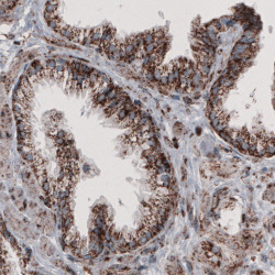
- Immunohistochemical staining of human prostate shows granular cytoplasmic immunoreactivity in glandular cells.

- Submitted by
- Atlas Antibodies (provider)
- Main image
- Experimental details
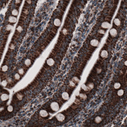
- Immunohistochemical staining of human small intestine shows strong granular cytoplasmic immunoreactivity in both glandular and lamina propria cells.

- Submitted by
- Atlas Antibodies (provider)
- Main image
- Experimental details
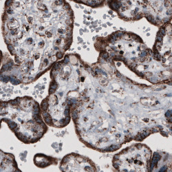
- Immunohistochemical staining of human placenta shows granular cytoplasmic positivity in the trophoblast.

- Submitted by
- Atlas Antibodies (provider)
- Main image

- Experimental details
- Immunohistochemical staining of human liver shows granular cytoplasmic positivity in hepatocytes.

- Submitted by
- Atlas Antibodies (provider)
- Main image
- Experimental details
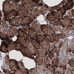
- Immunohistochemical staining of human skeletal muscle shows granular cytoplasmic immunoreactivity in muscle fibers.
