 Explore
 Validate
 Learn
Learn Western blot
 Immunoprecipitation
Antibody data
- Antibody Data
- Antigen structure
- References [1]
- Comments [0]
- Validations
- Western blot [11]
- Immunocytochemistry [4]
- Immunohistochemistry [8]
Submit
Validation data
Reference
Comment
Report error
- Product number
- PA5-22126 - Provider product page

- Provider
- Invitrogen Antibodies
- Product name
- Citrate Synthase Polyclonal Antibody
- Antibody type
- Polyclonal
- Antigen
- Recombinant protein fragment
- Description
- Recommended positive controls: 293T, A431, HeLa, HepG2, mouse brain, rat brain, Rat muscle. Predicted reactivity: Mouse (94%), Rat (93%), Zebrafish (90%), Xenopus laevis (91%), Pig (95%), Bovine (95%). Store product as a concentrated solution. Centrifuge briefly prior to opening the vial.
- Reactivity
- Human, Mouse, Rat, Chicken/Avian
- Host
- Rabbit
- Isotype
- IgG
- Vial size
- 100 µL
- Concentration
- 0.65 mg/mL
- Storage
- Store at 4°C short term. For long term storage, store at -20°C, avoiding freeze/thaw cycles.
Submitted references mRNA Therapy Improves Metabolic and Behavioral Abnormalities in a Murine Model of Citrin Deficiency.
Cao J, An D, Galduroz M, Zhuo J, Liang S, Eybye M, Frassetto A, Kuroda E, Funahashi A, Santana J, Mihai C, Benenato KE, Kumarasinghe ES, Sabnis S, Salerno T, Coughlan K, Miracco EJ, Levy B, Besin G, Schultz J, Lukacs C, Guey L, Finn P, Furukawa T, Giangrande PH, Saheki T, Martini PGV
Molecular therapy : the journal of the American Society of Gene Therapy 2019 Jul 3;27(7):1242-1251
Molecular therapy : the journal of the American Society of Gene Therapy 2019 Jul 3;27(7):1242-1251
No comments: Submit comment
Supportive validation
- Submitted by
- Invitrogen Antibodies (provider)
- Main image

- Experimental details
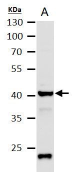
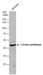
- Western blot analysis of Citrate synthase using 50µg of mouse brain lysate. Samples were loaded onto a 10% SDS-PAGE gel and probed with a Citrate synthase polyclonal antibody (Product # PA5-22126) at a dilution of 1:1000.
- Submitted by
- Invitrogen Antibodies (provider)
- Main image
- Experimental details
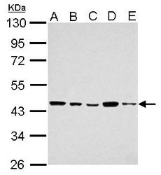
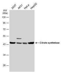
- Western blot analysis of Citrate synthase using A) 30 µg 293T whole cell extract (B) 30 µg A431 whole cell lysate (C) 30 µg HeLa whole cell lysate (D) 30 µg HepG2 whole cell lysate and E) 30 µg A375 whole cell lysate. Samples were loaded onto a 10% SDS-PAGE gel and probed with a Citrate synthase polyclonal antibody (Product # PA5-22126) at a dilution of 1:1000.
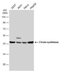
- Submitted by
- Invitrogen Antibodies (provider)
- Main image
- Experimental details
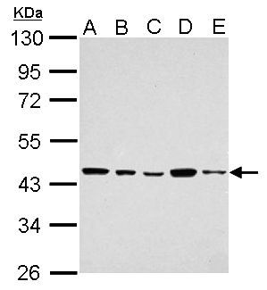
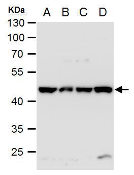
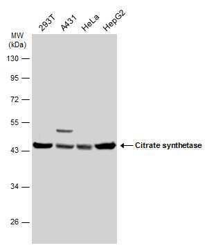
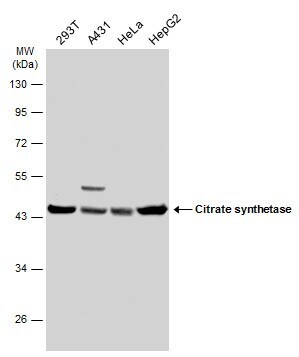
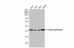
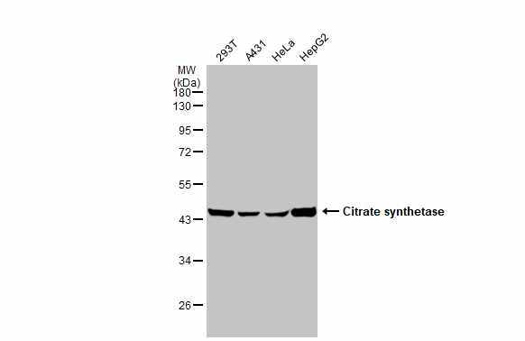
- Western blot analysis of Citrate synthase using A) 30 µg 293T whole cell extract (B) 30 µg A431 whole cell extract (C) 30 µg HeLa whole cell extract and D) 30 µg HepG2 whole cell extract. Samples were loaded onto a 10% SDS-PAGE gel and probed with a Citrate synthase polyclonal antibody (Product # PA5-22126) at a dilution of 1:1000.
- Submitted by
- Invitrogen Antibodies (provider)
- Main image
- Experimental details
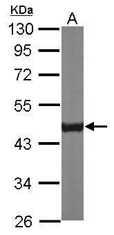
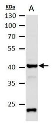
- Western blot analysis of Citrate synthase using 50 µg mouse brain extract. Samples were loaded onto a 10% SDS-PAGE gel and probed with a Citrate synthase polyclonal antibody (Product # PA5-22126) at a dilution of 1:1000.
- Submitted by
- Invitrogen Antibodies (provider)
- Main image
- Experimental details
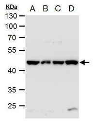
- Western Blot analysis of Citrate Synthase was performed by separating 30 µg of various whole cell extracts by 10% SDS-PAGE. Proteins were transferred to a membrane and probed with a Citrate Synthase Polyclonal Antibody (Product # PA5-22126) at a dilution of 1:1000 and a HRP-conjugated anti-rabbit IgG secondary antibody.
- Submitted by
- Invitrogen Antibodies (provider)
- Main image
- Experimental details
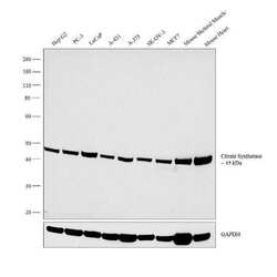
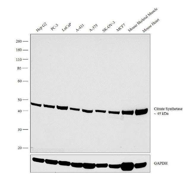
- Western blot analysis was performed on membrane enriched extracts (30 µg lysate) of Hep G2 (Lane 1), PC-3 (Lane 2), LnCaP (Lane 3), A-431 (Lane 4), A-375 (Lane 5), SK-OV-3 (Lane 6), MCF7 (Lane 7), tissue extracts of Mouse Skeletal Muscle (Lane 8) and Mouse Heart (Lane 9). The blot was probed with Anti-Citrate Synthetase Polyclonal Antibody (Product # PA5-22126, 1:2000 dilution) and detected by chemiluminescence using Goat anti-Rabbit IgG (H+L) Superclonal™ Secondary Antibody, HRP conjugate (Product # A27036, 0.25 µg/ml, 1:4000 dilution). A 45 kDa band corresponding to Citrate Synthetase was observed across the cell lines and tissues tested.
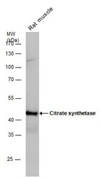
- Submitted by
- Invitrogen Antibodies (provider)
- Main image
- Experimental details
- Western Blot analysis of Citrate Synthase was performed by separating 50 µg of rat tissue extracts by 10% SDS-PAGE. Proteins were transferred to a membrane and probed with a Citrate Synthase Polyclonal Antibody (Product # PA5-22126) at a dilution of 1:1000. The HRP-conjugated anti-rabbit IgG antibody was used to detect the primary antibody.
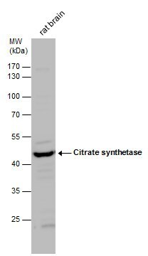
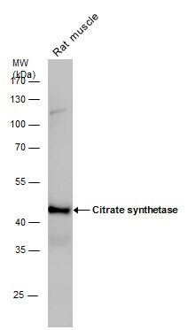
- Submitted by
- Invitrogen Antibodies (provider)
- Main image
- Experimental details
- Western Blot analysis of Citrate Synthase was performed by separating 50 µg of rat tissue extracts by 10% SDS-PAGE. Proteins were transferred to a membrane and probed with a Citrate Synthase Polyclonal Antibody (Product # PA5-22126) at a dilution of 1:1000.
- Submitted by
- Invitrogen Antibodies (provider)
- Main image
- Experimental details
- Western Blot analysis of Citrate Synthase was performed by separating 30 µg of various whole cell extracts by 10% SDS-PAGE. Proteins were transferred to a membrane and probed with a Citrate Synthase Polyclonal Antibody (Product # PA5-22126) at a dilution of 1:1000 and a HRP-conjugated anti-rabbit IgG secondary antibody.
- Submitted by
- Invitrogen Antibodies (provider)
- Main image
- Experimental details
- Western Blot using Citrate Synthase Polyclonal Antibody (Product # PA5-22126). Various whole cell extracts (30 µg) were separated by 10% SDS-PAGE, and the membrane was blotted with Citrate synthetase antibody [N2C3] Citrate Synthase Polyclonal Antibody (Product # PA5-22126) diluted at 1:1,000. The HRP-conjugated anti-rabbit IgG antibody was used to detect the primary antibody.
- Submitted by
- Invitrogen Antibodies (provider)
- Main image
- Experimental details
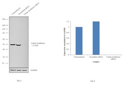
- Knockdown of Citrate Synthetase was achieved by transfecting Hep G2 with Citrate Synthetase specific siRNAs (Silencer® select Product # s3582). Western blot analysis (Fig. a) was performed using membrane enriched extracts from the Citrate Synthetase knockdown cells (lane 3), non-specific scrambled siRNA transfected cells (lane 2) and untransfected cells (lane 1). The blot was probed with Citrate Synthetase Polyclonal Antibody (Product # PA5-22126, 1:2000 dilution) and Goat anti-Rabbit IgG (H+L) Superclonal™ Secondary Antibody, HRP conjugate (Product # A27036, 0.25µg/ml, 1:4000 dilution). Densitometric analysis of this western blot is shown in histogram (Fig. b). Decrease in signal upon siRNA mediated knock down confirms that antibody is specific to Citrate Synthetase.

Supportive validation
- Submitted by
- Invitrogen Antibodies (provider)
- Main image

- Experimental details
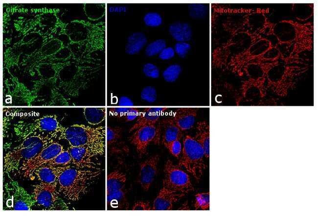
- Immunofluorescence analysis of Citrate synthase was performed using 70% confluent log phase Hep G2 cells. The cells were fixed with 4% paraformaldehyde for 10 minutes, permeabilized with 0.1% Triton™ X-100 for 15 minutes, and blocked with 1% BSA for 1 hour at room temperature. The cells were labeled with Citrate Synthetase Polyclonal Antibody (Product # PA5-22126) at 1:100 dilution in 0.1% BSA, incubated at 4 degree Celsius overnight and then labeled with Goat anti-Rabbit IgG (H+L) Superclonal™ Secondary Antibody, Alexa Fluor® 488 conjugate (Product # A27034) at a dilution of 1:2000 for 45 minutes at room temperature (Panel a: green). Nuclei (Panel b: blue) were stained with SlowFade® Gold Antifade Mountant with DAPI (Product # S36938). Mitochondria (Panel c: red) was stained with Mitotracker Red CMXRos (Product # M7512). Panel d represents the merged image showing mitochondrial localization. Panel e represents control cells with no primary antibody to assess background. The images were captured at 60X magnification.
- Submitted by
- Invitrogen Antibodies (provider)
- Main image

- Experimental details
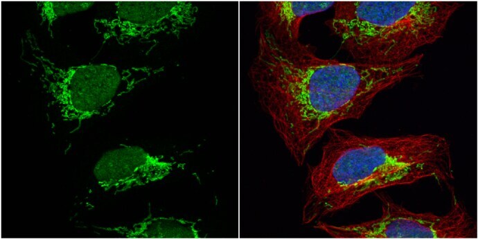
- Immunocytochemistry-Immunofluorescence analysis of Citrate Synthase was performed in U2OS cells fixed in 4% paraformaldehyde at RT for 15 min. Green: Citrate Synthase Polyclonal Antibody (Product # PA5 22126) diluted at 1:500. Red: alpha Tubulin, a cytoskeleton marker. Blue: Hoechst 33342 staining.
- Submitted by
- Invitrogen Antibodies (provider)
- Main image

- Experimental details
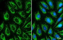
- Immunocytochemistry-Immunofluorescence analysis of Citrate Synthase was performed in HeLa cells fixed in 4% paraformaldehyde at RT for 15 min. Green: Citrate Synthase Polyclonal Antibody (Product # PA5 22126) diluted at 1:500. Blue: Hoechst 33342 staining. Scale bar = 10 µm.

- Submitted by
- Invitrogen Antibodies (provider)
- Main image
- Experimental details
- Citrate synthetase antibody [N2C3] detects Citrate synthetase protein at mitochondria by immunofluorescent analysis. Sample: HeLa cells were fixed in 4% paraformaldehyde at RT for 15 min. Green: Citrate synthetase stained by Citrate synthetase antibody [N2C3] (Product # PA5-22126) diluted at 1:500. Blue: Fluoroshield with DAPI . Scale bar= 10 μm.

Supportive validation
- Submitted by
- Invitrogen Antibodies (provider)
- Main image

- Experimental details
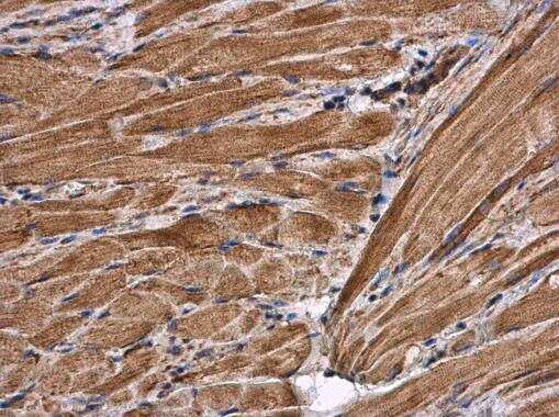
- Immunohistochemistry (Paraffin) analysis of Citrate Synthase was performed in paraffin-embedded mouse muscle tissue using Citrate Synthase Polyclonal Antibody (Product # PA5-22126) at a dilution of 1:500.
- Submitted by
- Invitrogen Antibodies (provider)
- Main image

- Experimental details
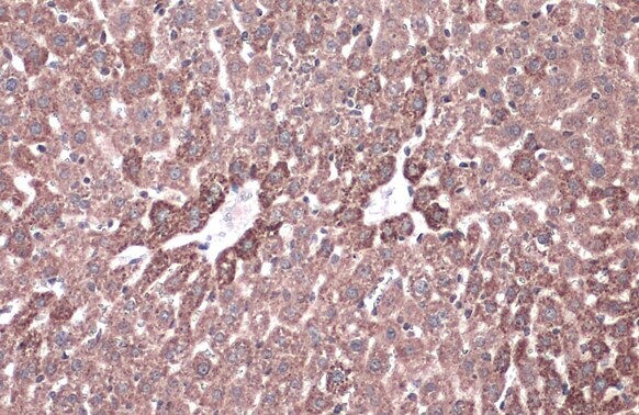
- Immunohistochemistry (Paraffin) analysis of Citrate Synthase was performed in paraffin-embedded mouse liver tissue using Citrate Synthase Polyclonal Antibody (Product # PA5-22126) at a dilution of 1:500. Antigen Retrieval: Citrate buffer, pH 6.0, 15 min.
- Submitted by
- Invitrogen Antibodies (provider)
- Main image
- Experimental details
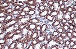
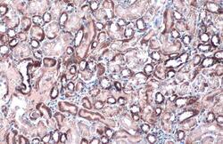
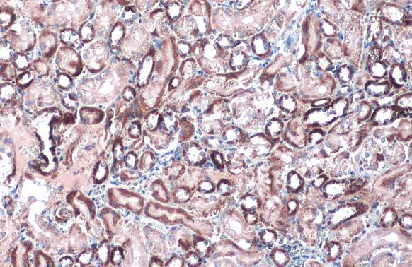
- Immunohistochemistry (Paraffin) analysis of Citrate Synthase was performed in paraffin-embedded rat kidney tissue using Citrate Synthase Polyclonal Antibody (Product # PA5-22126) at a dilution of 1:500. Antigen Retrieval: Citrate buffer, pH 6.0, 15 min.
- Submitted by
- Invitrogen Antibodies (provider)
- Main image
- Experimental details
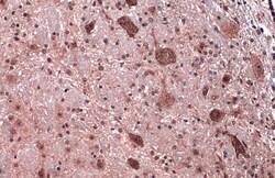
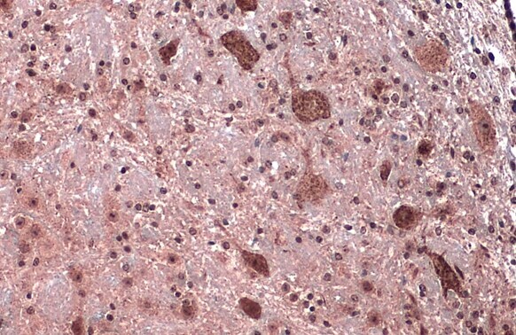
- Immunohistochemistry (Paraffin) analysis of Citrate Synthase was performed in paraffin-embedded mouse brain tissue using Citrate Synthase Polyclonal Antibody (Product # PA5-22126) at a dilution of 1:500. Antigen Retrieval: Citrate buffer, pH 6.0, 15 min.
- Submitted by
- Invitrogen Antibodies (provider)
- Main image
- Experimental details
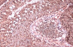
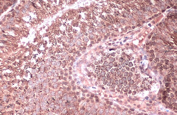
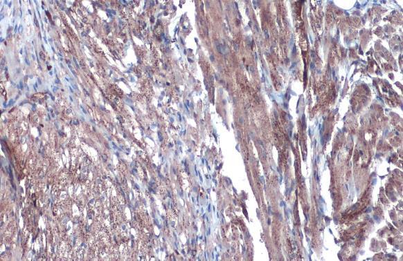
- Immunohistochemistry (Paraffin) analysis of Citrate Synthase was performed in paraffin-embedded rat testis tissue using Citrate Synthase Polyclonal Antibody (Product # PA5-22126) at a dilution of 1:500. Antigen Retrieval: Citrate buffer, pH 6.0, 15 min.
- Submitted by
- Invitrogen Antibodies (provider)
- Main image
- Experimental details
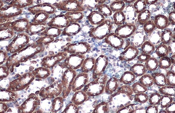
- Citrate synthetase antibody [N2C3] detects Citrate synthetase protein at mitochondria by immunohistochemical analysis. Sample: Paraffin-embedded mouse kidney. Citrate synthetase stained by Citrate synthetase antibody [N2C3] (Product # PA5-22126) diluted at 1:500. Antigen Retrieval: Citrate buffer, pH 6.0, 15 min.
- Submitted by
- Invitrogen Antibodies (provider)
- Main image

- Experimental details
- Citrate synthetase antibody [N2C3] detects Citrate synthetase protein at mitochondria by immunohistochemical analysis. Sample: Paraffin-embedded rat heart. Citrate synthetase stained by Citrate synthetase antibody [N2C3] (Product # PA5-22126) diluted at 1:500. Antigen Retrieval: Citrate buffer, pH 6.0, 15 min.
- Submitted by
- Invitrogen Antibodies (provider)
- Main image
- Experimental details
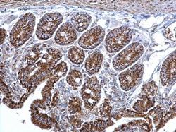
- Citrate synthetase antibody [N2C3] detects Citrate synthetase protein at mitochondria on mouse intestine by immunohistochemical analysis. Sample: Paraffin-embedded mouse intestine. Citrate synthetase antibody [N2C3] (Product # PA5-22126) dilution: 1:500. Antigen Retrieval: EDTA based buffer, pH 8.0, 15 min.
