 Explore
 Validate
 Learn
Learn Western blot
Antibody data
- Antibody Data
- Antigen structure
- References [0]
- Comments [0]
- Validations
- Western blot [2]
- ELISA [1]
- Immunohistochemistry [1]
Submit
Validation data
Reference
Comment
Report error
- Product number
- LS-C793551 - Provider product page

- Provider
- LSBio
- Product name
- DCLK3 / CLR Antibody (N-Terminus) LS-C793551
- Antibody type
- Polyclonal
- Description
- Affinity purified
- Reactivity
- Human
- Host
- Rabbit
- Isotype
- IgG
- Storage
- Store at -20°C. Avoid freeze-thaw cycles.
No comments: Submit comment
Supportive validation
- Submitted by
- LSBio (provider)
- Main image

- Experimental details
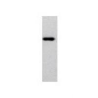
- Western Blot: DCLK3 Antibody - Western Blot was performed using affinity purified SEQer DCAMKL3 antibody, aa(138-237) antibody. The lanes contain 5-30ug of a whole cell extract. Final concentration of antibodies = 0.1ug/ml. (1:10,000 dilution). The blot was probed overnight with the SEQer DCAMKL3 antibody, aa(138-237) antibody. Blot was then washed according to protocol and probed with goat-anti-Rabbit-HRP conjugate at 1:5000 dilution, washed and developed using chemiluminescence. (film exposure 5-30sec). The protein was detected as represented by the band shown.
- Submitted by
- LSBio (provider)
- Main image

- Experimental details
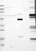
- Western Blot: DCLK3 Antibody - Samples: Lane 1, Marker: 250, 130, 95, 72, 55, 36, 28, 17, 11 Lane 2, RT-4 Lane 3, U-251MG sp Lane 4, Human Plasma Lane 5, Liver Lane 6, Tonsil , Target weight: 74 Validation score: 2 Validation description: Supportive - Band of predicted size in kDa. (+/-20%) with additional bands present.
Supportive validation
- Submitted by
- LSBio (provider)
- Main image
- Experimental details
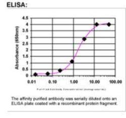
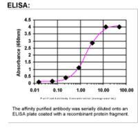
- ELISA: DCLK3 Antibody
Supportive validation
- Submitted by
- LSBio (provider)
- Main image
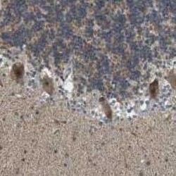
- Experimental details
- Immunohistochemical staining of human cerebellum shows cytoplasmic and nuclear positivity in Purkinje cells. Antibody dilution: 1:400. Image and statement courtesy of the Human Protein Atlas. (HPA).
