Explore
 Validate
 Learn
Learn Immunohistochemistry
Antibody data
- Antibody Data
- Antigen structure
- References [1]
- Comments [0]
- Validations
- Immunohistochemistry [1]
- Other assay [1]
Submit
Validation data
Reference
Comment
Report error
- Product number
- PA5-53106 - Provider product page

- Provider
- Invitrogen Antibodies
- Product name
- NEBL Polyclonal Antibody
- Antibody type
- Polyclonal
- Antigen
- Recombinant protein fragment
- Description
- Immunogen sequence: GQGIMNKEPA VIGRPDFEHA VEASKLSSQI KYKEKFDNEM KDKKHHYNPL ESASFRQNQL AATLASNVKY KKDIQNMHDP VSDLPNLLFL DHVLKASKML SGREYKKLFE ENKGMYHFDA DAVEHLHHKG NAVLQ Highest antigen sequence identity to the following orthologs: Mouse - 83%, Rat - 84%.
- Reactivity
- Human
- Host
- Rabbit
- Isotype
- IgG
- Vial size
- 100 μL
- Concentration
- 0.1 mg/mL
- Storage
- Store at 4°C short term. For long term storage, store at -20°C, avoiding freeze/thaw cycles.
Submitted references QKI is a critical pre-mRNA alternative splicing regulator of cardiac myofibrillogenesis and contractile function.
Chen X, Liu Y, Xu C, Ba L, Liu Z, Li X, Huang J, Simpson E, Gao H, Cao D, Sheng W, Qi H, Ji H, Sanderson M, Cai CL, Li X, Yang L, Na J, Yamamura K, Liu Y, Huang G, Shou W, Sun N
Nature communications 2021 Jan 4;12(1):89
Nature communications 2021 Jan 4;12(1):89
No comments: Submit comment
Supportive validation
- Submitted by
- Invitrogen Antibodies (provider)
- Main image
- Experimental details
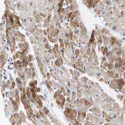
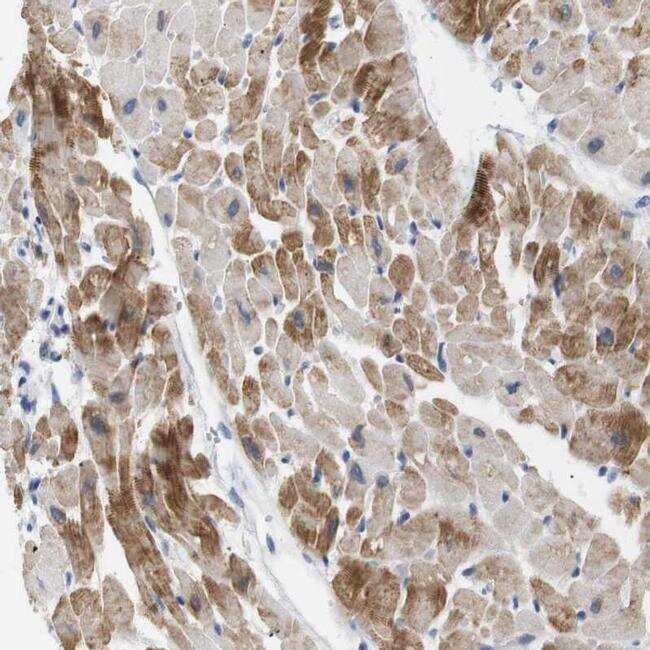
- Immunohistochemical analysis of NEBL in human heart muscle using NEBL Polyclonal Antibody (Product # PA5-53106) shows moderate cytoplasmic positivity in myocytes.
Supportive validation
- Submitted by
- Invitrogen Antibodies (provider)
- Main image
- Experimental details
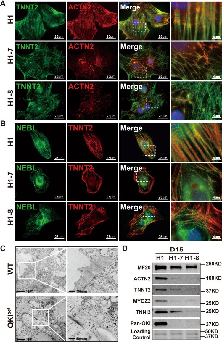
- Fig. 7 Severely altered myofibrillogenesis in hESCs- QKI del -derived cardiomyocytes. A Representative images of immunofluorescence staining of TNNT2 (green) and ACTN2 (red) in H1, H1-7, and H1-8 cardiomyocytes at differentiation Day-15. Scale bar: 25 mum. B Representative images of immunofluorescence staining of TNNT2 (red) and NEBL (green) in H1, H1-7, and H1-8 cardiomyocytes at differentiation Day-15. Scale bar: 25 mum. C Representative transmission electron microscopy images of normal and QKI-deficient differentiated cardiomyocytes at Day-15. Scale bar: 2 mum and 500 nm, respectively. D Western blot analysis of key myofibrillar proteins in control H1 and mutant H1-7 and H1-8 cardiomyocytes (Day-15). All experiments are independently repeated three times with three different sets of cell samples to ensure the reproducibility of the findings.