Explore
 Validate
 Learn
Learn Western blot
 Immunohistochemistry
Antibody data
- Antibody Data
- Antigen structure
- References [2]
- Comments [0]
- Validations
- Western blot [1]
Submit
Validation data
Reference
Comment
Report error
- Product number
- PB9204 - Provider product page

- Provider
- Boster Biological Technology
- Product name
- Anti-Glutamate Receptor 1/GRIA1 Antibody Picoband™
- Antibody type
- Polyclonal
- Description
- Polyclonal antibody for GluR1/GRIA1 detection. Host: Rabbit.Size: 100μg/vial. Tested applications: IHC-P. Reactive species: Human. GluR1/GRIA1 information: Molecular Weight: 101506 MW; Subcellular Localization: Cell membrane ; Multi-pass membrane protein . Endoplasmic reticulum membrane ; Multi-pass membrane protein . Cell junction, synapse, postsynaptic cell membrane ; Multi-pass membrane protein . Cell junction, synapse, postsynaptic cell membrane, postsynaptic density . Cell projection, dendrite . Cell projection, dendritic spine . Interaction with CACNG2, CNIH2 and CNIH3 promotes cell surface expression; Tissue Specificity: Widely expressed in brain.
- Reactivity
- Human, Mouse, Rat
- Host
- Rabbit
- Vial size
- 100μg/vial
- Concentration
- Add 0.2ml of distilled water will yield a concentration of 500ug/ml.
- Storage
- At -20°C for one year. After reconstitution, at 4°C for one month. It can also be aliquoted and stored frozen at -20°C for a longer time. Avoid repeated freezing and thawing.
- Handling
- Add 0.2ml of distilled water will yield a concentration of 500ug/ml.
Submitted references Disruption of glutamate neurotransmitter transmission is modulated by SNAP-25 in benzo[a]pyrene-induced neurotoxic effects.
Loss of the golgin GM130 causes Golgi disruption, Purkinje neuron loss, and ataxia in mice.
Yang K, Jiang X, Su Q, Wang J, Li C, Xia Y, Cheng S, Qin Q, Cao X, Chen C, Tu B
Toxicology 2017 Jun 1;384:11-22
Toxicology 2017 Jun 1;384:11-22
Loss of the golgin GM130 causes Golgi disruption, Purkinje neuron loss, and ataxia in mice.
Liu C, Mei M, Li Q, Roboti P, Pang Q, Ying Z, Gao F, Lowe M, Bao S
Proceedings of the National Academy of Sciences of the United States of America 2017 Jan 10;114(2):346-351
Proceedings of the National Academy of Sciences of the United States of America 2017 Jan 10;114(2):346-351
No comments: Submit comment
Supportive validation
- Submitted by
- Boster Biological Technology (provider)
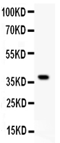
- Main image

- Experimental details
- Western blot analysis of GRIA1 using anti-GRIA1 antibody (PB9204). Electrophoresis was performed on a 5-20% SDS-PAGE gel at 70V (Stacking gel) / 90V (Resolving gel) for 2-3 hours. lane 1: Recombinant Human GRIA1 Protein 0.5ng. After Electrophoresis, proteins were transferred to a Nitrocellulose membrane at 150mA for 50-90 minutes. Blocked the membrane with 5% Non-fat Milk/ TBS for 1.5 hour at RT. The membrane was incubated with rabbit anti-GRIA1 antigen affinity purified polyclonal antibody (Catalog # PB9204) at 0.5 μg/mL overnight at 4°C, then washed with TBS-0.1%Tween 3 times with 5 minutes each and probed with a goat anti-rabbit IgG-HRP secondary antibody at a dilution of 1:10000 for 1.5 hour at RT. The signal is developed using an Enhanced Chemiluminescent detection (ECL) kit (Catalog # EK1002) with Tanon 5200 system. A specific band was detected for GRIA1 at approximately 40KD. The expected band size for GRIA1 is at 40KD.
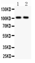
- Additional image