Explore
 Validate
 Learn
Learn Western blot
 Immunoelectron microscopy
Antibody data
- Antibody Data
- Antigen structure
- References [0]
- Comments [0]
- Validations
- Western blot [2]
- Immunocytochemistry [1]
- Immunohistochemistry [1]
Submit
Validation data
Reference
Comment
Report error
- Product number
- PA5-111797 - Provider product page

- Provider
- Invitrogen Antibodies
- Product name
- GluR1 (extracellular) Polyclonal Antibody
- Antibody type
- Polyclonal
- Antigen
- Synthetic peptide
- Description
- Applications Reported: This eBioCB16 (CB16) antibody has been reported for use in flow cytometric analysis.
- Reactivity
- Human, Mouse, Rat
- Host
- Guinea Pig
- Isotype
- IgG
- Vial size
- 50 µL
- Concentration
- 0.8 mg/mL
- Storage
- -20°C
No comments: Submit comment
Supportive validation
- Submitted by
- Invitrogen Antibodies (provider)
- Main image
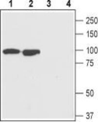
- Experimental details
- Western Blot analysis of GluR1 was performed in rat (lanes 1 and 3) and mouse (lanes 2 and 4) brain lysates. Lane 1,2: GluR1 (extracellular) Antibody (Product # PA5-111797) at a dilution of 1:200. Lane 3,4: GluR1 (extracellular) Antibody preincubated with the negative control antigen.

- Submitted by
- Invitrogen Antibodies (provider)
- Main image
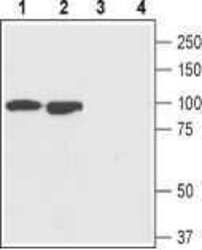
- Experimental details
- Western Blot analysis of GluR1 was performed in rat (lanes 1 and 3) and mouse (lanes 2 and 4) brain lysates. Lane 1,2: GluR1 (extracellular) Antibody (Product # PA5-111797) at a dilution of 1:200. Lane 3,4: GluR1 (extracellular) Antibody preincubated with the negative control antigen.

Supportive validation
- Submitted by
- Invitrogen Antibodies (provider)
- Main image

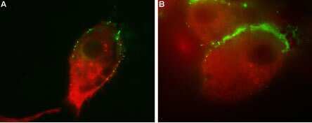
- Experimental details
- Immunocytochemistry-Immunofluorescence analysis of GluR1 in human U-87 MG cells using GluR1 (extracellular) Antibody (Product # PA5-111797), (1:25), followed by goat anti-guinea pig-AlexaFluor-488 secondary antibody (green). Cells were subsequently fixed, permeabilized and labeled with a GABA Transporter Antibody, followed by goat anti-rabbit-AlexaFluor-594 secondary antibody (red). Representative merged images of the double labeled cells are shown in A and B.
Supportive validation
- Submitted by
- Invitrogen Antibodies (provider)
- Main image
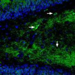
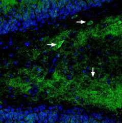
- Experimental details
- Immunohistochemistry analysis of GluR1 in perfusion-fixed, frozen rat hippocampus tissue sections using GluR1 (extracellular) Antibody (Product # PA5-111797) at a dilution of 1:400, followed by anti-rabbit-Cy2 antibody (green). GluR1 staining appears in neuronal outlines (horizontal arrows) and in the inner molecular layer of the dentate gyrus (vertical arrow). Nuclei are stained with DAPI (blue).