Explore
 Validate
 Learn
Learn Western blot
Antibody data
- Antibody Data
- Antigen structure
- References [1]
- Comments [0]
- Validations
- Western blot [2]
- Immunocytochemistry [2]
- Immunohistochemistry [3]
- Other assay [1]
Submit
Validation data
Reference
Comment
Report error
- Product number
- MA5-25530 - Provider product page

- Provider
- Invitrogen Antibodies
- Product name
- PNMT Monoclonal Antibody (OTI1D2)
- Antibody type
- Monoclonal
- Antigen
- Recombinant full-length protein
- Reactivity
- Human, Mouse, Rat, Canine
- Host
- Mouse
- Isotype
- IgG
- Antibody clone number
- OTI1D2
- Vial size
- 100 μL
- Concentration
- 1.0 mg/mL
- Storage
- -20°C, Avoid Freeze/Thaw Cycles
Submitted references β-Adrenoceptor Activation in Breast MCF-10A Cells Induces a Pattern of Catecholamine Production Similar to that of Tumorigenic MCF-7 Cells.
Amaro F, Silva D, Reguengo H, Oliveira JC, Quintas C, Vale N, Gonçalves J, Fresco P
International journal of molecular sciences 2020 Oct 27;21(21)
International journal of molecular sciences 2020 Oct 27;21(21)
No comments: Submit comment
Supportive validation
- Submitted by
- Invitrogen Antibodies (provider)
- Main image

- Experimental details
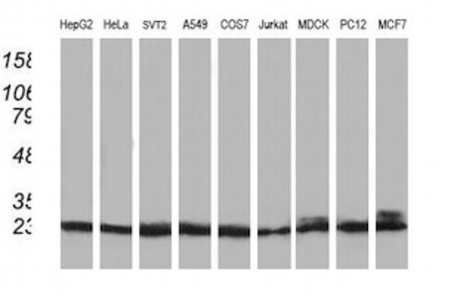
- Western blot analysis of PNMT in HepG2, HeLa, SVT2, A549, COS7, Jurkat, MDCK, PC12, MCF7 cells using 35 µg per lane. Samples were probed with PNMT (Product # MA5-25530) monoclonal antibody.
- Submitted by
- Invitrogen Antibodies (provider)
- Main image
- Experimental details
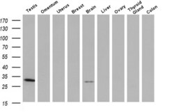
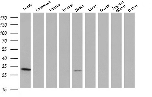
- Western blot analysis of PNMT in human tissue (1: Testis; 2: Omentum; 3: Uterus; 4: Breast; 5: Brain; 6: Liver; 7: Ovary; 8: Thyroid gland; 9: Colon) samples using 10 µg per lane. Samples were separated by SDS-PAGE and probed with PNMT (Product # MA5-25530) monoclonal antibody at a dilution of 1:200.
Supportive validation
- Submitted by
- Invitrogen Antibodies (provider)
- Main image

- Experimental details
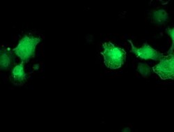
- Immunofluorescent analysis of PNMT in COS7 cells. Cells were transfected with a plasmid overexpressing PNMT and probed with a PNMT monoclonal antibody (Product # MA5-25530).

- Submitted by
- Invitrogen Antibodies (provider)
- Main image
- Experimental details
- Immunofluorescent analysis of PNMT in COS7 cells. Cells were transfected with a plasmid overexpressing PNMT and probed with a PNMT monoclonal antibody (Product # MA5-25530).

Supportive validation
- Submitted by
- Invitrogen Antibodies (provider)
- Main image
- Experimental details
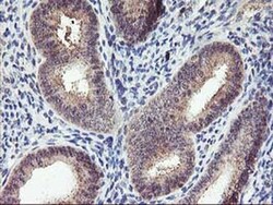
- Immunohistochemistry was performed on paraffin-embedded human endometrium tissue. To expose target proteins, 10mM citric buffer, pH6.0, 100°C for 10min was used. Following antigen retrieval, tissues were probed with a PNMT monoclonal antibody (Product # MA5-25530).
- Submitted by
- Invitrogen Antibodies (provider)
- Main image
- Experimental details
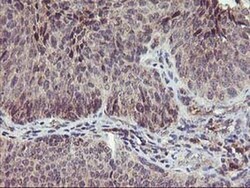
- Immunohistochemistry was performed on paraffin-embedded carcinoma of human bladder tissue. To expose target proteins, 10mM citric buffer, pH6.0, 100°C for 10min was used. Following antigen retrieval, tissues were probed with a PNMT monoclonal antibody (Product # MA5-25530).

- Submitted by
- Invitrogen Antibodies (provider)
- Main image
- Experimental details
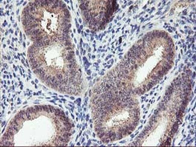
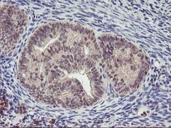
- Immunohistochemistry was performed on paraffin-embedded adenocarcinoma of human endometrium tissue. To expose target proteins, 10mM citric buffer, pH6.0, 100°C for 10min was used. Following antigen retrieval, tissues were probed with a PNMT monoclonal antibody (Product # MA5-25530).

Supportive validation
- Submitted by
- Invitrogen Antibodies (provider)
- Main image
- Experimental details
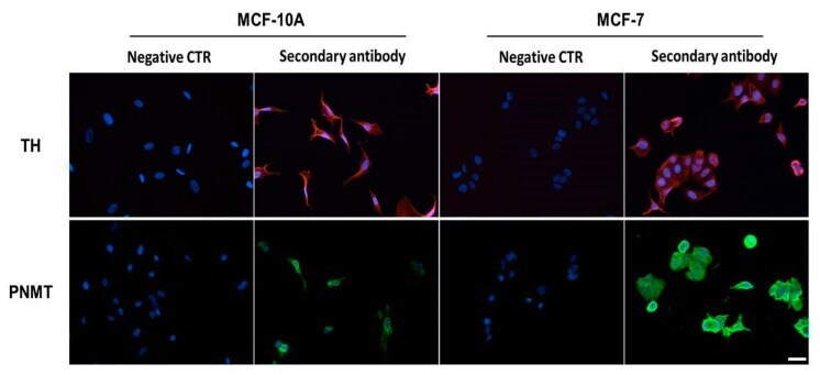
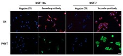
- Figure 2 Protein expression of tyrosine hydroxylase (TH) and phenylethanolamine N-methyltransferase (PNMT) in MCF-10A or in MCF-7 cells, revealed by immunocytochemistry. Shown are representative microphotographs of immunoreactivities revealed by secondary antibodies conjugated with Alexa fluor 594 (red fluorescence, TH) or with Alexa fluor 488 (green fluorescence, PNMT). Nuclei were labelled with Hoechst 33342 (blue fluorescence). Scale bar: 20 um. Negative control (CTR): without primary antibody.