Explore
 Validate
 Learn
Learn Western blot
 Immunoprecipitation
Antibody data
- Antibody Data
- Antigen structure
- References [0]
- Comments [0]
- Validations
- Western blot [1]
- Immunocytochemistry [1]
- Immunohistochemistry [1]
- Flow cytometry [1]
Submit
Validation data
Reference
Comment
Report error
- Product number
- GTX22814 - Provider product page

- Provider
- GeneTex
- Proper citation
- GeneTex Cat#GTX22814, RRID:AB_384869
- Product name
- p23 antibody [JJ3]
- Antibody type
- Monoclonal
- Reactivity
- Human, Mouse, Rat, Chicken/Avian, Guinea Pig, Rabbit
- Host
- Mouse
No comments: Submit comment
Supportive validation
- Submitted by
- GeneTex (provider)
- Main image
- Experimental details
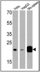
- Western blot analysis of p23 in 25 ug of HeLa, HepG2 and mouse spleen cell lysates. Proteins were transferred to a PVDF membrane and blocked with a blocking buffer at 4¢XC overnight. The membrane was probed with p23 antibody [JJ3] at a dilution of 1:1000 overnight at 4¢XC, washed in TBST, and probed with an HRP-conjugated secondary antibody. Chemiluminescent detection was performed.

Supportive validation
- Submitted by
- GeneTex (provider)
- Main image
- Experimental details
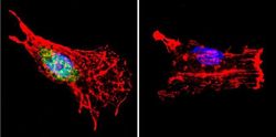
- Immunofluorescent analysis of p23 in C6 cells. p23 staining (green), F-Actin staining with Phalloidin (red) and nuclei with DAPI (blue) is shown. Cells were grown on slides and fixed with formaldehyde prior to staining. Cells were probed without (control) or with p23 antibody [JJ3] at a dilution of 1:500 over night at 4?C, washed with PBS and incubated with a proper secondary antibody. Images were taken at 60X magnification.

Supportive validation
- Submitted by
- GeneTex (provider)
- Main image
- Experimental details
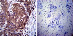
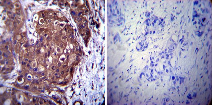
- Immunohistochemistry was performed on cancer biopsies of deparaffinized human breast carcinoma tissue. To expose target proteins, heat induced antigen retrieval was performed using 10mM sodium citrate (pH6.0) buffer, microwaved for 8-15 minutes. Following antigen retrieval tissues were blocked in 3% BSA-PBS for 30 minutes at room temperature. Tissues were then probed at a dilution of 1:1000 with or without p23 antibody [JJ3] overnight at 4¢XC in a humidified chamber. Tissues were washed extensively with PBST and endogenous peroxidase activity was quenched with a peroxidase suppressor. Detection was performed using a biotin-conjugated secondary antibody and SA-HRP, followed by colorimetric detection using DAB. Tissues were counterstained with hematoxylin and prepped for mounting.
Supportive validation
- Submitted by
- GeneTex (provider)
- Main image

- Experimental details
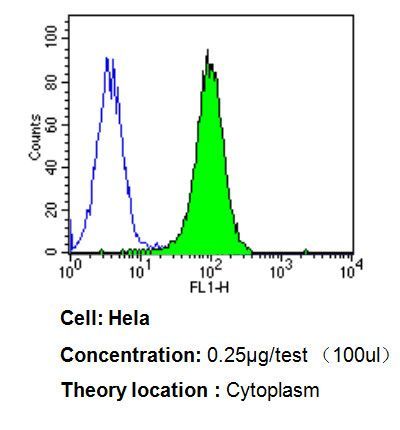
- Flow cytometry analysis of p23 in HeLa cells compared to an isotype control (blue). Cells were harvested, adjusted to a concentration of 1-5x10^6 cells/ml, fixed with 2% paraformaldehyde, washed with PBS, and incubated with p23 antibody [JJ3] at a dilution of 0.25 ug/test for 60 min at room temperature. Cells were then blocked in a solution of 2% BSA-PBS for 30 min at room temperature, incubated for 40 min at room temperature in the dark using a proper secondary antibody, and re-suspended in PBS for FACS analysis.