 Explore
 Validate
 Learn
Learn Western blot
Antibody data
- Antibody Data
- Antigen structure
- References [0]
- Comments [0]
- Validations
- Western blot [2]
- Immunocytochemistry [2]
- Other assay [1]
Submit
Validation data
Reference
Comment
Report error
- Product number
- 712913 - Provider product page

- Provider
- Invitrogen Antibodies
- Product name
- FTO Recombinant Superclonal™ Antibody (2HCLC)
- Antibody type
- Other
- Antigen
- Other
- Description
- This antibody is predicted to react with Mouse, Rat, Bovine, Recombinant rabbit Superclonal™ antibodies are unique offerings from Thermo Fisher Scientific. They are comprised of a selection of multiple different recombinant monoclonal antibodies, providing the best of both worlds - the sensitivity of polyclonal antibodies with the specificity of monoclonal antibodies - all delivered with the consistency only found in a recombinant antibody. While functionally the same as a polyclonal antibody - recognizing multiple epitope sites on the target and producing higher detection sensitivity for low abundance targets - a recombinant rabbit Superclonal™ antibody has a known mixture of light and heavy chains. The exact population can be produced in every lot, circumventing the biological variability typically associated with polyclonal antibody production. Note: Formerly called “Recombinant polyclonal antibody”, this product is now rebranded as “Recombinant Superclonal™ antibody”. The physical product and the performance remain unchanged.
- Reactivity
- Human
- Host
- Rabbit
- Isotype
- IgG
- Antibody clone number
- 2HCLC
- Vial size
- 100 μg
- Concentration
- 0.5 mg/mL
- Storage
- Store at 4°C short term. For long term storage, store at -20°C, avoiding freeze/thaw cycles.
No comments: Submit comment
Supportive validation
- Submitted by
- Invitrogen Antibodies (provider)
- Main image
- Experimental details
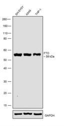
- Western blot was performed using Anti-FTO Rabbit Recombinant Rabbit Superclonal™ Antibody (Product # 712913) and a 58kDa band corresponding to FTO was observed across cell line tested. Whole cell extracts (30 µg lysate) of SH-SY5Y (Lane 1), A549 (Lane 2), and THP-1 (Lane 3) were electrophoresed using Novex® NuPAGE™ 4-12% Bis-Tris Protein Gel (Product # NP0321BOX). Resolved proteins were then transferred onto a nitrocellulose membrane (Product # IB23001) by iBlot® 2 Dry Blotting System (Product # IB21001). The blot was probed with the primary antibody (1:500 dilution) and detected by chemiluminescence Goat anti-Rabbit IgG (H+L) Superclonal™ Secondary Antibody, HRP conjugate (Product # A27036) at a 1:5000 dilution. Chemiluminescent detection was performed using Novex® ECL Chemiluminescent Substrate Reagent Kit (Product # WP20005).

- Submitted by
- Invitrogen Antibodies (provider)
- Main image
- Experimental details
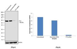
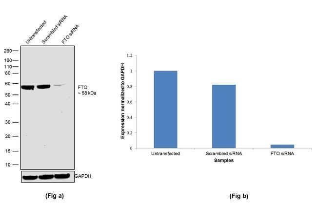
- Knockdown of FTO was achieved by transfecting A549 cells with FTO specific siRNA (Silencer® select Product # S35511, Product # S35512) . Western blot analysis (Fig a) was performed using whole cell extracts (1% SDS) from FTO knockdown cells (Lane 3), non-specific scrambled siRNA transfected cells (Lane 2) and untransfected cells (Lane 1). The blot was probed with Anti-FTO Recombinant Rabbit Superclonal™ Antibody (Product # 712913) at a 1:500 dilution and Goat anti-Rabbit IgG (Heavy Chain) Superclonal™ Secondary Antibody, HRP conjugate (Product # A27036) (0.25 µg/mL, 1:4000 dilution). Densitometric analysis of this western blot is shown in the histogram (Fig b). Loss of signal upon siRNA mediated knockdown confirms that antibody is specific to FTO.
Supportive validation
- Submitted by
- Invitrogen Antibodies (provider)
- Main image
- Experimental details
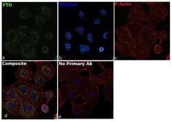
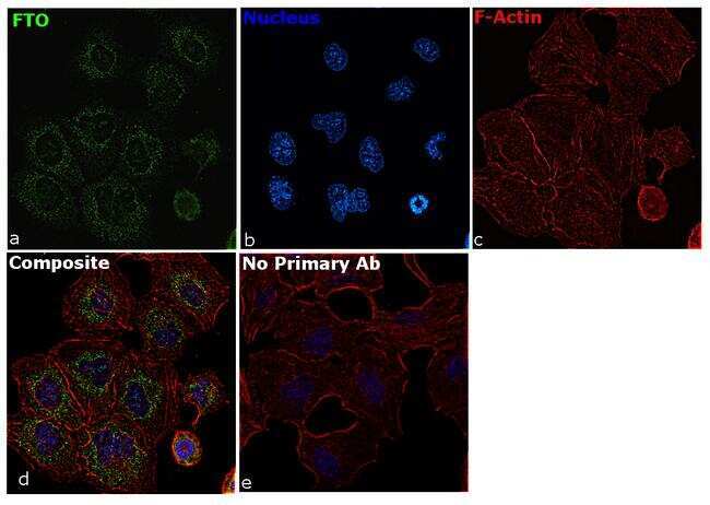
- For immunocytochemistry analysis, HeLa cells were fixed and permeabilized for detection of endogenous FTO using Anti-FTO Recombinant Rabbit Polycloclonal Antibody (Product # 712913) at a 1:100 dilution and labeled with Goat anti-Rabbit IgG (H+L) Superclonal™ Secondary Antibody, Alexa Fluor® 488 conjugate (Product # A27034) at a 1:2000 dilution. Panel a) shows representative cells that were stained for detection and localization of FTO protein (green), Panel b) is stained for nuclei (blue) using ProLong™ Diamond Antifade Mountant with DAPI (Product # P36962). Panel c) represents cytoskeletal F-actin staining using Rhodamine Phalloidin (Product # R415) at a 1:300 dilution. Panel d) is a composite image of Panels a, b and c clearly demonstrating cytoplasm and nuclear speckles localization of FTO. Panel e) represents control cells with no primary antibody to assess background. The images were captured at 60X magnification.
- Submitted by
- Invitrogen Antibodies (provider)
- Main image
- Experimental details
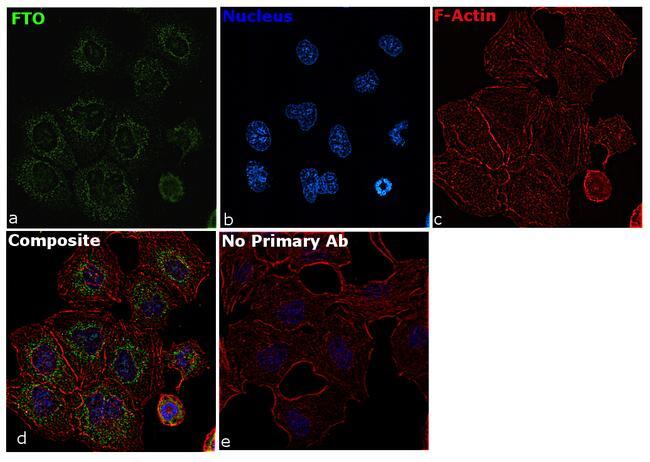
- For immunocytochemistry analysis, HeLa cells were fixed and permeabilized for detection of endogenous FTO using Anti-FTO Recombinant Rabbit Polycloclonal Antibody (Product # 712913) at a 1:100 dilution and labeled with Goat anti-Rabbit IgG (Heavy Chain) Superclonal™ Secondary Antibody, Alexa Fluor® 488 conjugate (Product # A27034) at a 1:2000 dilution. Panel a) shows representative cells that were stained for detection and localization of FTO protein (green), Panel b) is stained for nuclei (blue) using ProLong™ Diamond Antifade Mountant with DAPI (Product # P36962). Panel c) represents cytoskeletal F-actin staining using Rhodamine Phalloidin (Product # R415) at a 1:300 dilution. Panel d) is a composite image of Panels a, b and c clearly demonstrating cytoplasm and nuclear speckles localization of FTO. Panel e) represents control cells with no primary antibody to assess background. The images were captured at 60X magnification.
Supportive validation
- Submitted by
- Invitrogen Antibodies (provider)
- Main image
- Experimental details
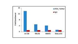
- Detection of binding of endogenous FTO protein to specific RNA using Anti-FTO Antibody: RNA Immunoprecipitation (RIP) was performed using 5 µg of Anti-FTO Recombinant Rabbit Polyclonal Antibody (Product # 712913) on whole cell lysate from 2 million SH-SY5Y cells. Normal Rabbit IgG was used as a negative IP control. RNA purified by RiboPure™ RNA Purification Kit (Product # AM1924) was analyzed by RT-PCR using the Power SYBR® Green RNA-to-CT™ 1-Step Kit (Product # 4389986) with RIP primer pairs over ACTB (positive), PIGM, SRSF2 mRNA and MALAT1 (negative). Data is presented as fold enrichment of the antibody signal versus the negative control IgG using the comparative CT method.
