Antibody data
- Antibody Data
- Antigen structure
- References [0]
- Comments [0]
- Validations
- Western blot [6]
- Immunocytochemistry [1]
- Other assay [1]
Submit
Validation data
Reference
Comment
Report error
- Product number
- PA5-78074 - Provider product page
- Provider
- Invitrogen Antibodies
- Product name
- NEK1 Polyclonal Antibody
- Antibody type
- Polyclonal
- Antigen
- Recombinant full-length protein
- Description
- Positive Control: 293T, mouse brain, rat brain Store product as a concentrated solution. Centrifuge briefly prior to opening the vial.
- Reactivity
- Human, Mouse
- Host
- Rabbit
- Isotype
- IgG
- Vial size
- 100 µL
- Concentration
- 0.33 mg/mL
- Storage
- Store at 4°C short term. For long term storage, store at -20°C, avoiding freeze/thaw cycles.
No comments: Submit comment
Supportive validation
- Submitted by
- Invitrogen Antibodies (provider)
- Main image
- Experimental details
- Western blot analysis of NEK1 in whole cell lysate using 30 µg of protein. Samples were separated with 5% SDS-PAGE and incubated with NEK1 polyclonal antibody (Product # PA5-78074) using a dilution of 1:1000.
- Submitted by
- Invitrogen Antibodies (provider)
- Main image
- Experimental details
- Western blot analysis of NEK1 in whole cell lysate using 50 µg of protein. Samples were separated with 5% SDS-PAGE and incubated with NEK1 polyclonal antibody (Product # PA5-78074) using a dilution of 1:1000.
- Submitted by
- Invitrogen Antibodies (provider)
- Main image
- Experimental details
- Western blot analysis of NEK1 was performed by loading 10 µg of WT (lane 1) and NEK1 CRISPR KO (lane 2) HeLa cell lysates in RIPA buffer onto a 4-15% gradient polyacrylamide gel. Proteins were transferred to nitrocellulose membrane and blocked in 5% milk. Ponceau stained transfer of blot is shown. NEK1 was detected using a NEK1 polyclonal antibody (Product # PA5-78074) at a dilution of 1:1,000 in 5% BSA in TBST overnight at 4 deg, followed by secondary antibody diluted to 0.2 µg/mL using Goat anti-Rabbit IgG (H+L) HRP antibody (Product # 65-6120). Chemiluminescent detection was performed using Pierce ECL Western Blotting Substrate (Product # 32106). Data courtesy of YCharOS Inc., an open science company with the mission of characterizing commercially available antibodies using knockout validation.
- Submitted by
- Invitrogen Antibodies (provider)
- Main image
- Experimental details
- Western blot analysis of NEK1 was performed by separating 50 µg of various tissue extracts by 5% SDS-PAGE. Proteins were transferred to a membrane and probed with a NEK1 Polyclonal Antibody (Product # PA5-78074) at a dilution of 1:1000. The HRP-conjugated anti-rabbit IgG antibody was used to detect the primary antibody.
- Submitted by
- Invitrogen Antibodies (provider)
- Main image
- Experimental details
- Western Blot using NEK1 Polyclonal Antibody (Product # PA5-78074). Whole cell extract (30 µg) was separated by 5% SDS-PAGE, and the membrane was blotted with NEK1 Polyclonal Antibody (Product # PA5-78074) diluted at 1:1,000.
- Submitted by
- Invitrogen Antibodies (provider)
- Main image
- Experimental details
- Western Blot using NEK1 Polyclonal Antibody (Product # PA5-78074). Various tissue extracts (50 µg) were separated by 5% SDS-PAGE, and the membrane was blotted with NEK1 Polyclonal Antibody (Product # PA5-78074) diluted at 1:1,000.
Supportive validation
- Submitted by
- Invitrogen Antibodies (provider)
- Main image
- Experimental details
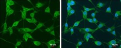
- NEK1 Polyclonal Antibody detects Nek1 protein at cytoplasm and nucleus by immunofluorescent analysis. Sample: U87-MG cells were fixed in 4% paraformaldehyde at RT for 15 min. Green: Nek1 protein stained by NEK1 Polyclonal Antibody (Product # PA5-78074) diluted at 1:500. Blue: Hoechst 33342 staining. Scale bar = 10 µm.
Supportive validation
- Submitted by
- Invitrogen Antibodies (provider)
- Main image
- Experimental details
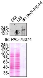
- Immunoprecipitation of NEK1 was performed on HeLa cell lysates. Antibody-bead conjugates were prepared by adding 1 µg of NEK1 polyclonal antibody (Product # PA5-78074) with 30 µL of protein A -Sepharose beads and rocked overnight at 4 deg. 1 µg of lysate was incubated with antibody-bead conjugate for 2 hrs at 4 deg. After multiple washes, 10% starting material (SM), 10% unbound fraction (UB) and immunoprecipitated fraction (IP) were processed for immunoblot using the same NEK1 antibody (Product # PA5-78074). Ponceau stained transfer of blot is shown. Data courtesy of YCharOS Inc., an open science company with the mission of characterizing commercially available antibodies using knockout validation.