Explore
 Validate
 Learn
Learn Western blot
 ELISA
ELISA Immunocytochemistry
Antibody data
- Antibody Data
- Antigen structure
- References [13]
- Comments [0]
- Validations
- Immunocytochemistry [2]
- Immunohistochemistry [3]
- Other assay [11]
Submit
Validation data
Reference
Comment
Report error
- Product number
- 33-8700 - Provider product page

- Provider
- Invitrogen Antibodies
- Product name
- p38 MAPK beta Monoclonal Antibody (P38-11A5)
- Antibody type
- Monoclonal
- Antigen
- Recombinant full-length protein
- Description
- 33-8700 has been successfully used in immunofluorescence and western blot. 33-8700 specifically recognizes the beta isoform of p38, with slight reactivity to p38-gamma.
- Reactivity
- Human, Mouse
- Host
- Mouse
- Isotype
- IgG
- Antibody clone number
- P38-11A5
- Vial size
- 100 μg
- Concentration
- 0.5 mg/mL
- Storage
- -20°C
Submitted references HuR stabilizes HTT mRNA via interacting with its exon 11 in a mutant HTT-dependent manner.
Discovery of a Small Molecule Promoting Mouse and Human Osteoblast Differentiation via Activation of p38 MAPK-β.
Loss of Functionally Redundant p38 Isoforms in T Cells Enhances Regulatory T Cell Induction.
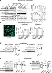
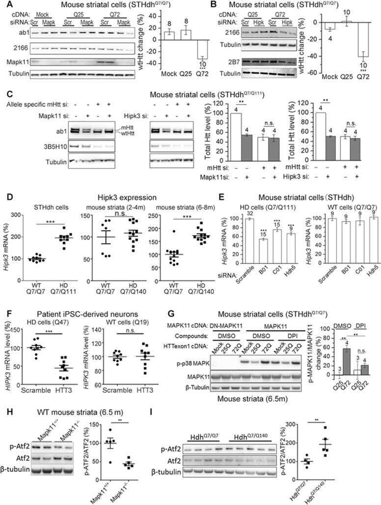
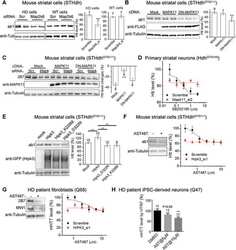
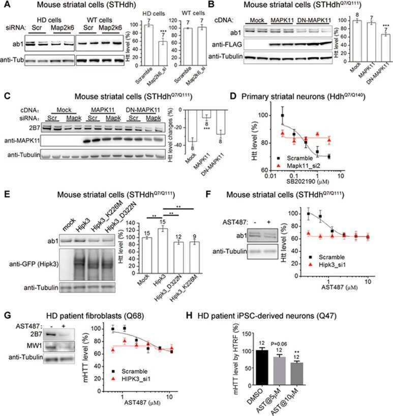
Suppression of MAPK11 or HIPK3 reduces mutant Huntingtin levels in Huntington's disease models.
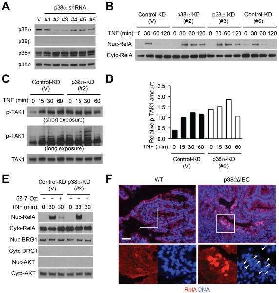
Epithelial Control of Gut-Associated Lymphoid Tissue Formation through p38α-Dependent Restraint of NF-κB Signaling.
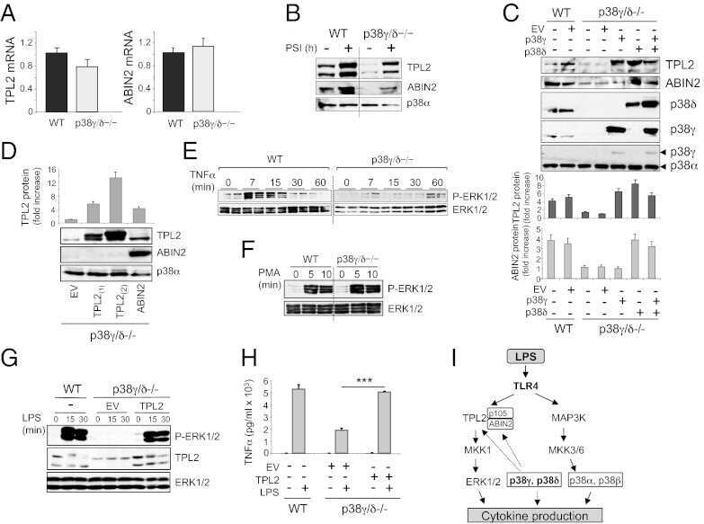
p38γ and p38δ kinases regulate the Toll-like receptor 4 (TLR4)-induced cytokine production by controlling ERK1/2 protein kinase pathway activation.
Cyclosporine A induces nerve growth factor expression via activation of MAPK p38 and NFAT5.
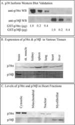
Pomegranate extract inhibits the interleukin-1β-induced activation of MKK-3, p38α-MAPK and transcription factor RUNX-2 in human osteoarthritis chondrocytes.
BIRB796 inhibits all p38 MAPK isoforms in vitro and in vivo.
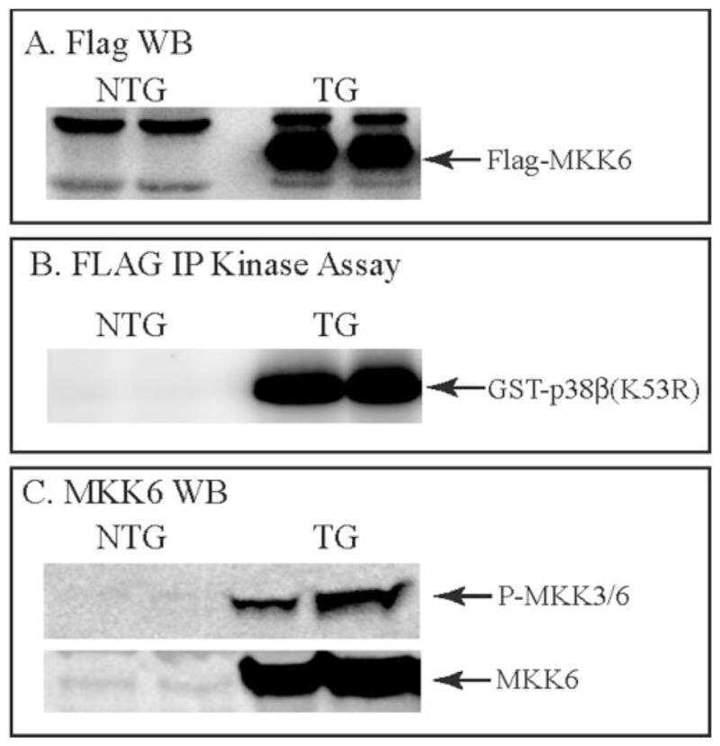
Overexpression of mitogen-activated protein kinase kinase 6 in the heart improves functional recovery from ischemia in vitro and protects against myocardial infarction in vivo.
Basic fibroblast growth factor-induced cell death is effected through sustained activation of p38MAPK and up-regulation of the death receptor p75NTR.
Stress kinase phosphorylation is increased in pacing-induced heart failure in rabbits.
Cooperation of p38 and extracellular signal-regulated kinase mitogen-activated protein kinase pathways during granulocyte colony-stimulating factor-induced hemopoietic cell proliferation.
Zhao Q, Li C, Yu M, Sun Y, Wang J, Ma L, Sun X, Lu B
RNA biology 2020 Apr;17(4):500-516
RNA biology 2020 Apr;17(4):500-516
Discovery of a Small Molecule Promoting Mouse and Human Osteoblast Differentiation via Activation of p38 MAPK-β.
Cook B, Rafiq R, Lee H, Banks KM, El-Debs M, Chiaravalli J, Glickman JF, Das BC, Chen S, Evans T
Cell chemical biology 2019 Jul 18;26(7):926-935.e6
Cell chemical biology 2019 Jul 18;26(7):926-935.e6
Loss of Functionally Redundant p38 Isoforms in T Cells Enhances Regulatory T Cell Induction.
Hayakawa M, Hayakawa H, Petrova T, Ritprajak P, Sutavani RV, Jiménez-Andrade GY, Sano Y, Choo MK, Seavitt J, Venigalla RKC, Otsu K, Georgopoulos K, Arthur JSC, Park JM
The Journal of biological chemistry 2017 Feb 3;292(5):1762-1772
The Journal of biological chemistry 2017 Feb 3;292(5):1762-1772
Suppression of MAPK11 or HIPK3 reduces mutant Huntingtin levels in Huntington's disease models.
Yu M, Fu Y, Liang Y, Song H, Yao Y, Wu P, Yao Y, Pan Y, Wen X, Ma L, Hexige S, Ding Y, Luo S, Lu B
Cell research 2017 Dec;27(12):1441-1465
Cell research 2017 Dec;27(12):1441-1465
Epithelial Control of Gut-Associated Lymphoid Tissue Formation through p38α-Dependent Restraint of NF-κB Signaling.
Caballero-Franco C, Guma M, Choo MK, Sano Y, Enzler T, Karin M, Mizoguchi A, Park JM
Journal of immunology (Baltimore, Md. : 1950) 2016 Mar 1;196(5):2368-76
Journal of immunology (Baltimore, Md. : 1950) 2016 Mar 1;196(5):2368-76
p38γ and p38δ kinases regulate the Toll-like receptor 4 (TLR4)-induced cytokine production by controlling ERK1/2 protein kinase pathway activation.
Risco A, del Fresno C, Mambol A, Alsina-Beauchamp D, MacKenzie KF, Yang HT, Barber DF, Morcelle C, Arthur JS, Ley SC, Ardavin C, Cuenda A
Proceedings of the National Academy of Sciences of the United States of America 2012 Jul 10;109(28):11200-5
Proceedings of the National Academy of Sciences of the United States of America 2012 Jul 10;109(28):11200-5
Cyclosporine A induces nerve growth factor expression via activation of MAPK p38 and NFAT5.
Lee JH, Kim JW, Im YS, Seong GJ, Lee HK
Cornea 2011 Oct;30 Suppl 1:S19-24
Cornea 2011 Oct;30 Suppl 1:S19-24
Pomegranate extract inhibits the interleukin-1β-induced activation of MKK-3, p38α-MAPK and transcription factor RUNX-2 in human osteoarthritis chondrocytes.
Rasheed Z, Akhtar N, Haqqi TM
Arthritis research & therapy 2010;12(5):R195
Arthritis research & therapy 2010;12(5):R195
BIRB796 inhibits all p38 MAPK isoforms in vitro and in vivo.
Kuma Y, Sabio G, Bain J, Shpiro N, Márquez R, Cuenda A
The Journal of biological chemistry 2005 May 20;280(20):19472-9
The Journal of biological chemistry 2005 May 20;280(20):19472-9
Overexpression of mitogen-activated protein kinase kinase 6 in the heart improves functional recovery from ischemia in vitro and protects against myocardial infarction in vivo.
Martindale JJ, Wall JA, Martinez-Longoria DM, Aryal P, Rockman HA, Guo Y, Bolli R, Glembotski CC
The Journal of biological chemistry 2005 Jan 7;280(1):669-76
The Journal of biological chemistry 2005 Jan 7;280(1):669-76
Basic fibroblast growth factor-induced cell death is effected through sustained activation of p38MAPK and up-regulation of the death receptor p75NTR.
Williamson AJ, Dibling BC, Boyne JR, Selby P, Burchill SA
The Journal of biological chemistry 2004 Nov 12;279(46):47912-28
The Journal of biological chemistry 2004 Nov 12;279(46):47912-28
Stress kinase phosphorylation is increased in pacing-induced heart failure in rabbits.
Schulz R, Aker S, Belosjorow S, Konietzka I, Rauen U, Heusch G
American journal of physiology. Heart and circulatory physiology 2003 Nov;285(5):H2084-90
American journal of physiology. Heart and circulatory physiology 2003 Nov;285(5):H2084-90
Cooperation of p38 and extracellular signal-regulated kinase mitogen-activated protein kinase pathways during granulocyte colony-stimulating factor-induced hemopoietic cell proliferation.
Rausch O, Marshall CJ
The Journal of biological chemistry 1999 Feb 12;274(7):4096-105
The Journal of biological chemistry 1999 Feb 12;274(7):4096-105
No comments: Submit comment
Supportive validation
- Submitted by
- Invitrogen Antibodies (provider)
- Main image

- Experimental details
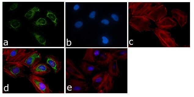
- Immunofluorescent analysis of p38-beta 2 MAPK Antibody (P38-11A5) was done on 70% confluent log phase HeLa cells. The cells were fixed with 4% paraformaldehyde for 15 minutes, permeabilized with 0.25% Triton™ X-100 for 10 minutes, and blocked with 5% BSA for 1 hour at room temperature. The cells were labeled with p38-beta 2 MAPK Antibody (P38-11A5) (Product # 33-8700) at 1µg/mL in 1% BSA and incubated for 3 hours at room temperature and then labeled with Alexa Fluor 488 Rabbit Anti-Mouse IgG Secondary Antibody (Product # A-11059) at a dilution of 1:400 for 45 minutes at room temperature (Panel a: green). Nuclei (Panel b: blue) were stained with SlowFade® Gold Antifade Mountant with DAPI (Product # S36938). F-actin (Panel c: red) was stained with Alexa Fluor 594 Phalloidin (Product # A12381). Panel d is a merged image showing cytoplasmic localization. Panel e is a no primary antibody control. The images were captured at 40X magnification.
- Submitted by
- Invitrogen Antibodies (provider)
- Main image

- Experimental details
- Immunofluorescent analysis of p38-beta 2 MAPK Antibody (P38-11A5) was done on 70% confluent log phase HeLa cells. The cells were fixed with 4% paraformaldehyde for 15 minutes, permeabilized with 0.25% Triton™ X-100 for 10 minutes, and blocked with 5% BSA for 1 hour at room temperature. The cells were labeled with p38-beta 2 MAPK Antibody (P38-11A5) (Product # 33-8700) at 1µg/mL in 1% BSA and incubated for 3 hours at room temperature and then labeled with Alexa Fluor 488 Rabbit Anti-Mouse IgG Secondary Antibody (Product # A-11059) at a dilution of 1:400 for 45 minutes at room temperature (Panel a: green). Nuclei (Panel b: blue) were stained with SlowFade® Gold Antifade Mountant with DAPI (Product # S36938). F-actin (Panel c: red) was stained with Alexa Fluor 594 Phalloidin (Product # A12381). Panel d is a merged image showing cytoplasmic localization. Panel e is a no primary antibody control. The images were captured at 40X magnification.

Supportive validation
- Submitted by
- Invitrogen Antibodies (provider)
- Main image

- Experimental details
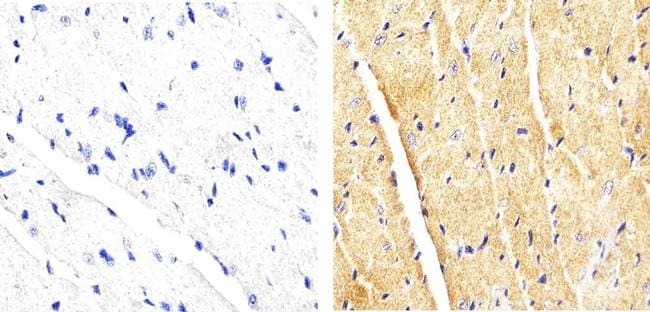
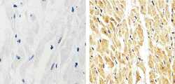
- Immunohistochemistry analysis of p38-beta2 MAPK showing staining in the cytoplasm and nucleus of paraffin-embedded mouse heart tissue (right) compared to a negative control without primary antibody (left). To expose target proteins, antigen retrieval was performed using 10mM sodium citrate (pH 6.0), microwaved for 8-15 min. Following antigen retrieval, tissues were blocked in 3% H2O2-methanol for 15 min at room temperature, washed with ddH2O and PBS, and then probed with a p38-beta2 MAPK monoclonal antibody (Product # 33-8700) diluted in 3% BSA-PBS at a dilution of 1:20 overnight at 4ºC in a humidified chamber. Tissues were washed extensively in PBST and detection was performed using an HRP-conjugated secondary antibody followed by colorimetric detection using a DAB kit. Tissues were counterstained with hematoxylin and dehydrated with ethanol and xylene to prep for mounting.
- Submitted by
- Invitrogen Antibodies (provider)
- Main image
- Experimental details
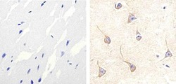
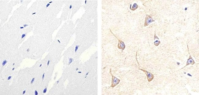
- Immunohistochemistry analysis of p38-beta2 MAPK showing staining in the cytoplasm and nucleus of paraffin-embedded human brain tissue (right) compared to a negative control without primary antibody (left). To expose target proteins, antigen retrieval was performed using 10mM sodium citrate (pH 6.0), microwaved for 8-15 min. Following antigen retrieval, tissues were blocked in 3% H2O2-methanol for 15 min at room temperature, washed with ddH2O and PBS, and then probed with a p38-beta2 MAPK monoclonal antibody (Product # 33-8700) diluted in 3% BSA-PBS at a dilution of 1:20 overnight at 4ºC in a humidified chamber. Tissues were washed extensively in PBST and detection was performed using an HRP-conjugated secondary antibody followed by colorimetric detection using a DAB kit. Tissues were counterstained with hematoxylin and dehydrated with ethanol and xylene to prep for mounting.
- Submitted by
- Invitrogen Antibodies (provider)
- Main image
- Experimental details
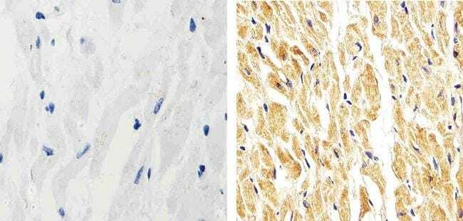
- Immunohistochemistry analysis of p38-beta2 MAPK showing staining in the cytoplasm and nucleus of paraffin-embedded human heart tissue (right) compared to a negative control without primary antibody (left). To expose target proteins, antigen retrieval was performed using 10mM sodium citrate (pH 6.0), microwaved for 8-15 min. Following antigen retrieval, tissues were blocked in 3% H2O2-methanol for 15 min at room temperature, washed with ddH2O and PBS, and then probed with a p38-beta2 MAPK monoclonal antibody (Product # 33-8700) diluted in 3% BSA-PBS at a dilution of 1:20 overnight at 4ºC in a humidified chamber. Tissues were washed extensively in PBST and detection was performed using an HRP-conjugated secondary antibody followed by colorimetric detection using a DAB kit. Tissues were counterstained with hematoxylin and dehydrated with ethanol and xylene to prep for mounting.
Supportive validation
- Submitted by
- Invitrogen Antibodies (provider)
- Main image
- Experimental details
- NULL
- Submitted by
- Invitrogen Antibodies (provider)
- Main image
- Experimental details
- NULL
- Submitted by
- Invitrogen Antibodies (provider)
- Main image
- Experimental details
- NULL
- Submitted by
- Invitrogen Antibodies (provider)
- Main image

- Experimental details
- NULL
- Submitted by
- Invitrogen Antibodies (provider)
- Main image
- Experimental details
- NULL

- Submitted by
- Invitrogen Antibodies (provider)
- Main image

- Experimental details
- NULL
- Submitted by
- Invitrogen Antibodies (provider)
- Main image
- Experimental details
- NULL
- Submitted by
- Invitrogen Antibodies (provider)
- Main image

- Experimental details
- NULL
- Submitted by
- Invitrogen Antibodies (provider)
- Main image
- Experimental details
- NULL
- Submitted by
- Invitrogen Antibodies (provider)
- Main image
- Experimental details
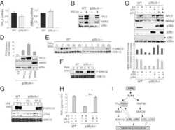
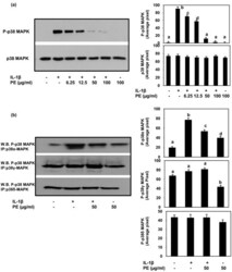
- Figure 4 Inhibition of IL-1beta-stimulated phosphorylation of p38-MAPK by PE in primary human OA chondrocytes . (a) Primary human OA chondrocytes were pretreated with PE for 2 h and then stimulated with IL-1beta for 30 minutes and total cell lysate proteins were resolved by SDS-PAGE and analyzed by Western immunoblotting using primary antibodies specific for phospho-p38-MAPK and total p38-MAPK. (b) Inhibition of IL-1beta-stimulated phosphorylation of p38-MAPK isoforms by PE in primary human OA chondrocytes. Primary human OA chondrocytes were pretreated with PE for 2 h and then stimulated with IL-1beta for 30 minutes and total cell lysate were used to immunoprecipitate p38-alpha, gamma and delta -MAPKs using monoclonal antibodies specific for each isoform as described above. Immunoprecipited protein was resolved by SDS-PAGE and Western blots were probed with anti-phospho-p38-MAPKantibody. Band images were digitally captured and the band intensities (pixels/band) were obtained as described under Figure 1. Average pixel values presented as mean +- SD; data without a common letter differ, P < 0.05.

- Submitted by
- Invitrogen Antibodies (provider)
- Main image

- Experimental details
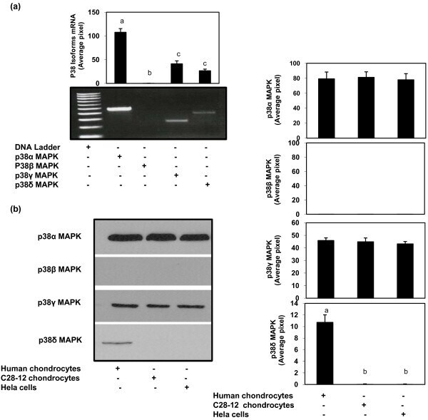
- Figure 3 Gene expression of p38-MAPK isoforms in primary human OA chondrocytes . (a) Total RNA was prepared from the human OA chondrocytes, single stranded cDNA was synthesized and the PCR reaction was carried out using specific primers for p38-MAPKalpha, beta, gamma and delta isoforms as described in methods section above. (b) Protein expression of p38-MAPK isoforms in human OA chondrocytes, C28-I2 human chondrocytes and Hela cells. Western blot analysis was performed as described under Figure 1. Band images were digitally captured and the band intensities (pixels/band) were obtained using the Un-Scan-It software. Data shown are cumulative of two experiments. Average pixel values presented as Mean +- SD; data without a common letter differ, P < 0.05.