Explore
 Validate
 Learn
Learn Western blot
 Immunohistochemistry
Antibody data
- Antibody Data
- Antigen structure
- References [0]
- Comments [0]
- Validations
- Western blot [1]
Submit
Validation data
Reference
Comment
Report error
- Product number
- PB9111 - Provider product page

- Provider
- Boster Biological Technology
- Product name
- Anti-Kv2.1/KCNB1 Antibody Picoband™
- Antibody type
- Polyclonal
- Description
- Polyclonal antibody for KV2.1/KCNB1 detection. Host: Rabbit.Size: 100μg/vial. Tested applications: IHC-P. Reactive species: Human. KV2.1/KCNB1 information: Molecular Weight: 95878 MW; Subcellular Localization: Cell membrane . Perikaryon . Cell projection, axon . Cell projection, dendrite . Membrane; Multi-pass membrane protein. Cell junction, synapse, postsynaptic cell membrane . Cell junction, synapse . Cell junction, synapse, synaptosome . Lateral cell membrane . Cell membrane, sarcolemma . Localizes to high-density somatodendritic clusters and non-clustered sites on the surface of neocortical and hippocampal pyramidal neurons in a cortical actin cytoskeleton-dependent manner (PubMed:24477962). Localizes also to high-density clusters in the axon initial segment (AIS), at ankyrin-G-deficient sites, on the surface of neocortical and hippocampal pyramidal neurons (PubMed:24477962). KCNB1-containing AIS clusters localize either in close apposition to smooth endoplasmic reticulum cisternal organelles or with GABA-A receptor-containing synapses of hippocampal and cortical pyramidal neurons, respectively (PubMed:24477962). Localizes to high-density clusters on the cell surface of atrial and ventricular myocytes and at the lateral plasma membrane in epithelial cells. Localizes both to the axial and transverse tubules (T tubule) and sarcolemma in ventricular myocytes. Associated with lipid raft domains. In cortical neurons, apoptotic injuries induce de novo plasma membrane insertion in a SNARE-dependent manner causing an apoptotic potassium current surge; Tissue Specificity: Expressed in neocortical pyramidal cells (PubMed:24477962). Expressed in pancreatic beta cells (at protein level) (PubMed:12403834, PubMed:14988243). Expressed in brain, heart, lung, liver, colon, kidney and adrenal gland (PubMed:19074135). Expressed in the cortex, amygdala, cerebellum, pons, thalamus, hypothalamus, hippocampus and substantia nigra (PubMed:19074135).
- Reactivity
- Human, Mouse, Rat
- Host
- Rabbit
- Vial size
- 100μg/vial
- Concentration
- Add 0.2ml of distilled water will yield a concentration of 500ug/ml.
- Storage
- At -20°C for one year. After reconstitution, at 4°C for one month. It can also be aliquoted and stored frozen at -20°C for a longer time. Avoid repeated freezing and thawing.
- Handling
- Add 0.2ml of distilled water will yield a concentration of 500ug/ml.
No comments: Submit comment
Supportive validation
- Submitted by
- Boster Biological Technology (provider)
- Main image
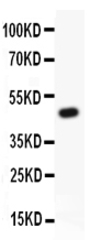
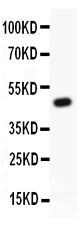
- Experimental details
- Western blot analysis of KV2.1 using anti-KV2.1 antibody (PB9111). Electrophoresis was performed on a 5-20% SDS-PAGE gel at 70V (Stacking gel) / 90V (Resolving gel) for 2-3 hours. lane 1:Recombinant Human kv2.1 Protein 0.5ng. After Electrophoresis, proteins were transferred to a Nitrocellulose membrane at 150mA for 50-90 minutes. Blocked the membrane with 5% Non-fat Milk/ TBS for 1.5 hour at RT. The membrane was incubated with rabbit anti-KV2.1 antigen affinity purified polyclonal antibody (Catalog # PB9111) at 0.5 μg/mL overnight at 4°C, then washed with TBS-0.1%Tween 3 times with 5 minutes each and probed with a goat anti-rabbit IgG-HRP secondary antibody at a dilution of 1:10000 for 1.5 hour at RT. The signal is developed using an Enhanced Chemiluminescent detection (ECL) kit (Catalog # EK1002) with Tanon 5200 system. A specific band was detected for KV2.1 at approximately 47KD. The expected band size for KV2.1 is at 47KD.
- Additional image