 Explore
 Validate
 Learn
Learn Western blot
Antibody data
- Antibody Data
- Antigen structure
- References [1]
- Comments [0]
- Validations
- Western blot [1]
- Flow cytometry [1]
Submit
Validation data
Reference
Comment
Report error
- Product number
- MAB11795 - Provider product page

- Provider
- Novus Biologicals
- Product name
- Rat Monoclonal AGER Antibody
- Antibody type
- Monoclonal
- Description
- Protein A or G purified from hybridoma culture supernatant. Detects mouse RAGE in direct ELISAs and Western blots. In Western blots, approximately 15% cross-reactivity with recombinant canine RAGE and no cross-reactivity with recombinant human RAGE or recombinant rat RAGE is observed.
- Reactivity
- Mouse
- Host
- Rat
- Conjugate
- Unconjugated
- Isotype
- IgG
- Vial size
- 100 ug
- Concentration
- LYOPH
- Storage
- Use a manual defrost freezer and avoid repeated freeze-thaw cycles. 12 months from date of receipt, -20 to -70 degreesC as supplied. 1 month, 2 to 8 degreesC under sterile conditions after reconstitution. 6 months, -20 to -70 degreesC under sterile conditions after reconstitution.
Submitted references Serum developmental endothelial locus-1 is associated with severity of sepsis in animals and humans.
Kim WY, Lee SH, Kim DY, Ryu HJ, Chon GR, Park YY, Fu Y, Huh JW, Lim CM, Koh Y, Choi EY, Hong SB
Scientific reports 2019 Sep 10;9(1):13005
Scientific reports 2019 Sep 10;9(1):13005
No comments: Submit comment
Supportive validation
- Submitted by
- Novus Biologicals (provider)
- Main image
- Experimental details
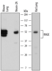
- Detection of Mouse and Rat RAGE by Western Blot. Western blot shows lysates of mouse lung tissue, Neuro-2A mouse neuroblastoma cell line, and rat lung tissue. PVDF membrane was probed with 1 µg/mL of Rat Anti-Mouse RAGE Monoclonal Antibody (Catalog # MAB11795) followed by HRP-conjugated Anti-Rat IgG Secondary Antibody (Catalog # HAF005). Specific bands were detected for RAGE at approximately 45 to 50 kDa (as indicated). This experiment was conducted under reducing conditions and using Immunoblot Buffer Group 1.

Supportive validation
- Submitted by
- Novus Biologicals (provider)
- Main image

- Experimental details
- Detection of RAGE in Mouse Splenocytes by Flow Cytometry. Mouse splenocytes stimulated to induce Th1 cells were stained with Rat Anti-Mouse CD4 PE-conjugated Monoclonal Antibody (Catalog # FAB554P) and either (A) Rat Anti-Mouse RAGE Monoclonal Antibody (Catalog # MAB11795) or (B) Rat IgG2A Isotype Control (Catalog # MAB006) followed by Allophycocyanin-conjugated Anti-Rat IgG Secondary Antibody (Catalog # F0113). View our protocol for Staining Membrane-associated Proteins.
