Explore
 Validate
 Learn
Learn43-8900
antibody from Invitrogen Antibodies
Targeting: NDUFA13
B16.6, CDA016, CGI-39, GRIM-19, GRIM19
 Western blot
 Immunocytochemistry
Antibody data
- Antibody Data
- Antigen structure
- References [2]
- Comments [0]
- Validations
- Immunocytochemistry [3]
- Flow cytometry [1]
- Other assay [2]
Submit
Validation data
Reference
Comment
Report error
- Product number
- 43-8900 - Provider product page

- Provider
- Invitrogen Antibodies
- Product name
- NDUFA13 Monoclonal Antibody (6E1BH7)
- Antibody type
- Monoclonal
- Antigen
- Recombinant full-length protein
- Description
- Positive control: Human heart, Bovine heart, Rat heart, and Mouse heart isolated mitochondria, Human fibroblasts, HeLa cells
- Reactivity
- Human, Mouse, Rat, Bovine
- Host
- Mouse
- Isotype
- IgG
- Antibody clone number
- 6E1BH7
- Vial size
- 100 μg
- Concentration
- 1 mg/mL
- Storage
- 4°C, do not freeze
Submitted references GDNF family ligand dependent STAT3 activation is mediated by specific alternatively spliced isoforms of GFRα2 and RET.
Mitochondrial localized STAT3 is involved in NGF induced neurite outgrowth.
Zhou L, Too HP
Biochimica et biophysica acta 2013 Dec;1833(12):2789-2802
Biochimica et biophysica acta 2013 Dec;1833(12):2789-2802
Mitochondrial localized STAT3 is involved in NGF induced neurite outgrowth.
Zhou L, Too HP
PloS one 2011;6(6):e21680
PloS one 2011;6(6):e21680
No comments: Submit comment
Supportive validation
- Submitted by
- Invitrogen Antibodies (provider)
- Main image
- Experimental details
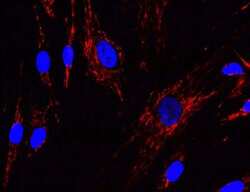
- Immunocytochemistry/Immunofluorescence analysis of GRIM19 in cultured human fibroblasts using a NDUFA13 Monoclonal Antibody (Product # 43-8900) at a concentration of 1 µg/mL. Cultured Human fibroblasts were fixed, permeabilized and then detected by Alexa® 488 goat-anti-mouse IgG.
- Submitted by
- Invitrogen Antibodies (provider)
- Main image

- Experimental details
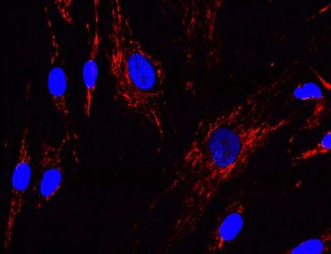
- Immunocytochemistry/Immunofluorescence analysis of GRIM19 in cultured human fibroblasts using a NDUFA13 Monoclonal Antibody (Product # 43-8900) at a concentration of 1 µg/mL. Cultured Human fibroblasts were fixed, permeabilized and then detected by Alexa® 488 goat-anti-mouse IgG.

- Submitted by
- Invitrogen Antibodies (provider)
- Main image
- Experimental details
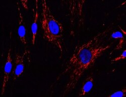
- Immunocytochemistry/Immunofluorescence analysis of GRIM19 in cultured human fibroblasts using a NDUFA13 Monoclonal Antibody (Product # 43-8900) at a concentration of 1 µg/mL. Cultured Human fibroblasts were fixed, permeabilized and then detected by Alexa® 488 goat-anti-mouse IgG.

Supportive validation
- Submitted by
- Invitrogen Antibodies (provider)
- Main image
- Experimental details
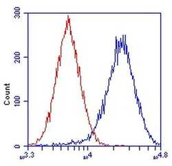
- Flow cytometric analysis of NDUFA13 in HeLa cells using a NDUFA13 Monoclonal Antibody (Product # 43-8900) at 1 µg/mL, as shown in blue. Isotype control antibody is shown in red.

Supportive validation
- Submitted by
- Invitrogen Antibodies (provider)
- Main image
- Experimental details
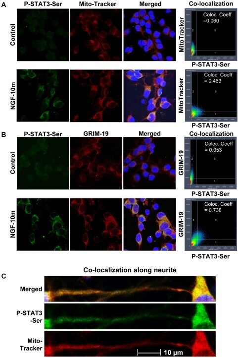
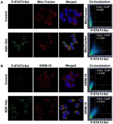
- Figure 5 P-Ser-STAT3 was co-localized with MitoTracker and GRIM-19 in PC12 cells. NGF stimulated (10 min) PC12 cells were co-stained for P-Ser-STAT3 & MitoTracker ( A ) or P-Ser-STAT3 & GRIM-19 ( B ). Confocal images of control and NGF stimulated cells of the individual and merged channels are shown. Also shown here are the intensity correlations and co-localization coefficients between P-Ser-STAT3 and MitoTracker or GRIM-19.

- Submitted by
- Invitrogen Antibodies (provider)
- Main image
- Experimental details
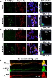
- Figure 6 P-Ser-STAT3 was co-localized with MitoTracker and GRIM-19 in rat embryonic cortical neuron. NGF stimulated (10 min) cortical neurons were co-stained for P-Ser-STAT3 & MitoTracker ( A, C ) or P-Ser-STAT3 & GRIM-19 ( B ). Confocal images of control and NGF stimulated cells of the individual and merged channels are shown. Also shown here are the intensity correlations and co-localization coefficients between P-Ser-STAT3 and MitoTracker or GRIM-19.