Explore
 Validate
 Learn
LearnHPA046717
antibody from Atlas Antibodies
Targeting: CES1
CEH, CES1A1, CES1A2, CES2, HMSE, HMSE1, SES1
 Immunocytochemistry
Antibody data
- Antibody Data
- Antigen structure
- References [1]
- Comments [0]
- Validations
- Immunocytochemistry [1]
- Immunohistochemistry [1]
Submit
Validation data
Reference
Comment
Report error
- Product number
- HPA046717 - Provider product page

- Provider
- Atlas Antibodies
- Proper citation
- Atlas Antibodies Cat#HPA046717, RRID:AB_2679770
- Product name
- Anti-CES1
- Antibody type
- Polyclonal
- Description
- Polyclonal Antibody against Human CES1, Gene description: carboxylesterase 1, Alternative Gene Names: CEH, CES1A1, CES1A2, CES2, HMSE, HMSE1, SES1, Validated applications: ICC, IHC, Uniprot ID: P23141, Storage: Store at +4°C for short term storage. Long time storage is recommended at -20°C.
- Reactivity
- Human
- Host
- Rabbit
- Conjugate
- Unconjugated
- Isotype
- IgG
- Vial size
- 100 µl
- Concentration
- 0.2 mg/ml
- Storage
- Store at +4°C for short term storage. Long time storage is recommended at -20°C.
- Handling
- The antibody solution should be gently mixed before use.
Submitted references Advantages of the Parent Nucleoside GS-441524 over Remdesivir for Covid-19 Treatment
Yan V, Muller F
ACS Medicinal Chemistry Letters 2020;11(7):1361-1366
ACS Medicinal Chemistry Letters 2020;11(7):1361-1366
No comments: Submit comment
Supportive validation
- Submitted by
- Atlas Antibodies (provider)
- Main image
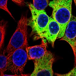
- Experimental details
- Immunofluorescent staining of human cell line Hep G2 shows localization to endoplasmic reticulum.
- Sample type
- Human

Supportive validation
- Submitted by
- Atlas Antibodies (provider)
- Enhanced method
- Orthogonal validation
- Main image

- Experimental details
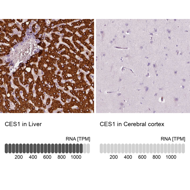
- Immunohistochemistry analysis in human liver and cerebral cortex tissues using HPA046717 antibody. Corresponding CES1 RNA-seq data are presented for the same tissues.
- Sample type
- Human
- Protocol
- Protocol