 Explore
 Validate
 Learn
Learn Western blot
 ELISA
ELISAAntibody data
- Antibody Data
- Antigen structure
- References [7]
- Comments [0]
- Validations
- Western blot [1]
- Immunocytochemistry [1]
- Immunoprecipitation [1]
- Immunohistochemistry [2]
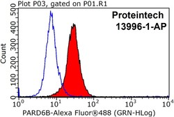
- Flow cytometry [1]
Submit
Validation data
Reference
Comment
Report error
- Product number
- 13996-1-AP - Provider product page

- Provider
- Proteintech Group
- Proper citation
- Proteintech Cat#13996-1-AP, RRID:AB_2159779
- Product name
- PARD6B antibody
- Antibody type
- Polyclonal
- Description
- PARD6B antibody (Cat. #13996-1-AP) is a rabbit polyclonal antibody that shows reactivity with human, mouse, rat and has been validated for the following applications: FC, IF, IHC, IP, WB,ELISA.
- Reactivity
- Human, Mouse, Rat
- Host
- Rabbit
- Conjugate
- Unconjugated
- Isotype
- IgG
- Vial size
- 20ul, 150ul
Submitted references Discovery and Functional Characterization of a Recombinant Fragment of Human Collagen Type XVII.
The PAR6B-PRKCI-PAR3 complex influences alveolar regeneration in patients with the emphysema subtype of chronic obstructive pulmonary disease.
PARD6A promotes lung adenocarcinoma cell proliferation and invasion through Serpina3.
Oncogenic functions and therapeutic potentials of targeted inhibition of SMARCAL1 in small cell lung cancer.
Induced retinal pigment epithelial cells with anti-epithelial-to-mesenchymal transition ability delay retinal degeneration.
PKCζ and JNK signaling regulate radiation-induced compensatory proliferation in parotid salivary glands.
Influence of Wilms' tumor suppressor gene WT1 on bovine Sertoli cells polarity and tight junctions via non-canonical WNT signaling pathway.
Piao L, Li J, Li X, Su Y, Yuan X, Chang S, Cheng X, Fu S, Kong R
Journal of agricultural and food chemistry 2025 Mar 19;73(11):6724-6735
Journal of agricultural and food chemistry 2025 Mar 19;73(11):6724-6735
The PAR6B-PRKCI-PAR3 complex influences alveolar regeneration in patients with the emphysema subtype of chronic obstructive pulmonary disease.
Wang D, Liu H, Bai S, Zheng X, Zhao L
Stem cell research & therapy 2025 Feb 25;16(1):97
Stem cell research & therapy 2025 Feb 25;16(1):97
PARD6A promotes lung adenocarcinoma cell proliferation and invasion through Serpina3.
Hu L, Liu M, Tang B, Li X, Xu H, Wang H, Wang D, Liu S, Xu C
Cancer gene therapy 2024 Nov;31(11):1696-1707
Cancer gene therapy 2024 Nov;31(11):1696-1707
Oncogenic functions and therapeutic potentials of targeted inhibition of SMARCAL1 in small cell lung cancer.
Sun BB, Wang GZ, Han SC, Yang FY, Guo H, Liu J, Liu YT, Zhou GB
Cancer letters 2024 Jun 28;592:216929
Cancer letters 2024 Jun 28;592:216929
Induced retinal pigment epithelial cells with anti-epithelial-to-mesenchymal transition ability delay retinal degeneration.
Tian H, Chen Z, Zhu X, Ou Q, Wang Z, Wu B, Xu JY, Jin C, Gao F, Wang J, Zhang J, Zhang J, Lu L, Xu GT
iScience 2022 Oct 21;25(10):105050
iScience 2022 Oct 21;25(10):105050
PKCζ and JNK signaling regulate radiation-induced compensatory proliferation in parotid salivary glands.
Wong WY, Allie S, Limesand KH
PloS one 2019;14(7):e0219572
PloS one 2019;14(7):e0219572
Influence of Wilms' tumor suppressor gene WT1 on bovine Sertoli cells polarity and tight junctions via non-canonical WNT signaling pathway.
Wang X, Adegoke EO, Ma M, Huang F, Zhang H, Adeniran SO, Zheng P, Zhang G
Theriogenology 2019 Oct 15;138:84-93
Theriogenology 2019 Oct 15;138:84-93
No comments: Submit comment
Supportive validation
- Submitted by
- Proteintech Group (provider)
- Main image
- Experimental details
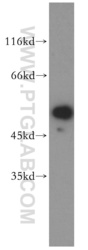
- HeLa cells were subjected to SDS PAGE followed by western blot with 13996-1-AP(PARD6B antibody) at dilution of 1:500
- Sample type
- cell line

Supportive validation
- Submitted by
- Proteintech Group (provider)
- Main image
- Experimental details
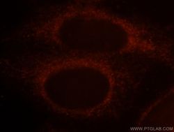
- Immunofluorescent analysis of HepG2 cells, using PARD6B antibody 13996-1-AP at 1:25 dilution and Rhodamine-labeled goat anti-rabbit IgG (red).
- Sample type
- cell line

Supportive validation
- Submitted by
- Proteintech Group (provider)
- Main image
- Experimental details
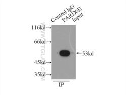
- IP Result of anti-PARD6B (IP:13996-1-AP, 3ug; Detection:13996-1-AP 1:200) with HEK-293 cells lysate 6000ug.
- Sample type
- cell line

Supportive validation
- Submitted by
- Proteintech Group (provider)
- Main image
- Experimental details
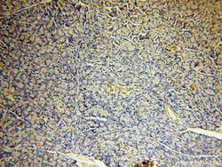
- Immunohistochemical of paraffin-embedded human pancreas using 13996-1-AP(PARD6B antibody) at dilution of 1:100 (under 10x lens)
- Sample type
- tissue

- Submitted by
- Proteintech Group (provider)
- Main image

- Experimental details
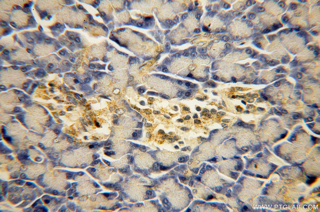
- Immunohistochemical of paraffin-embedded human pancreas using 13996-1-AP(PARD6B antibody) at dilution of 1:100 (under 40x lens)
- Sample type
- tissue
Supportive validation
- Submitted by
- Proteintech Group (provider)
- Main image
- Experimental details
- The PARD6B antibody from Proteintech is a rabbit polyclonal antibody to a recombinant protein of human PARD6B. This antibody recognizes human,mouse,rat antigen. The PARD6B antibody has been validated for the following applications: ELISA, WB, IHC, IF, IP, FC analysis.
