Explore
 Validate
 Learn
Learn Western blot
 Immunocytochemistry
Antibody data
- Antibody Data
- Antigen structure
- References [0]
- Comments [0]
- Validations
- Immunocytochemistry [2]
- Immunohistochemistry [4]
Submit
Validation data
Reference
Comment
Report error
- Product number
- PA5-53050 - Provider product page

- Provider
- Invitrogen Antibodies
- Product name
- PARD6B Polyclonal Antibody
- Antibody type
- Polyclonal
- Antigen
- Recombinant protein fragment
- Description
- Immunogen sequence: NYHKAVSTAN PLLRIFIQKK EEADYSAFGT DTLIKKKNVL TNVLRPDNHR KKPHIVISMP QDFRPVSSII DVDILPETHR RV Highest antigen sequence identity to the following orthologs: Mouse - 96%, Rat - 96%.
- Reactivity
- Human
- Host
- Rabbit
- Isotype
- IgG
- Vial size
- 100 μL
- Concentration
- 0.10 mg/mL
- Storage
- Store at 4°C short term. For long term storage, store at -20°C, avoiding freeze/thaw cycles.
No comments: Submit comment
Supportive validation
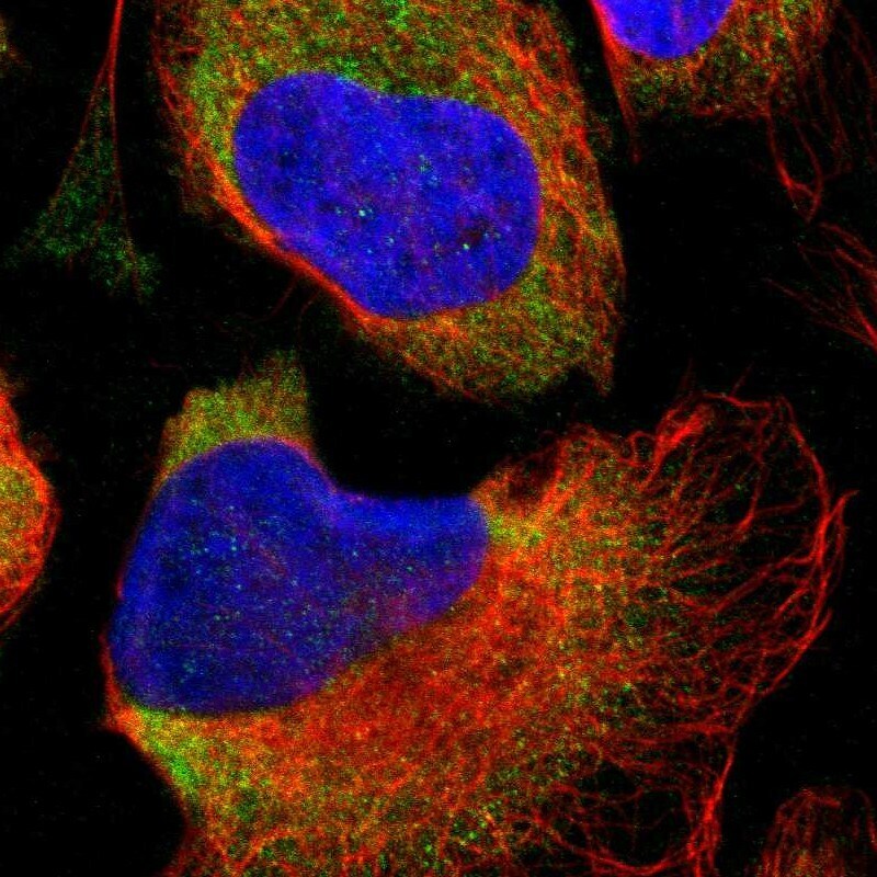
- Submitted by
- Invitrogen Antibodies (provider)
- Main image

- Experimental details
- Immunofluorescent staining of PARD6B in human cell line U-2 OS shows positivity in cytoplasm. Samples were probed using a PARD6B Polyclonal Antibody (Product # PA5-53050).
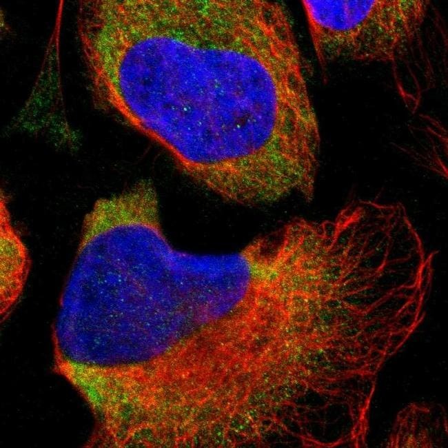
- Submitted by
- Invitrogen Antibodies (provider)
- Main image

- Experimental details
- Immunofluorecent analysis of PARD6B in human cell line U-2 OS using PARD6B Polyclonal Antibody (Product # PA5-53050). Staining shows localization to cytosol.
Supportive validation
- Submitted by
- Invitrogen Antibodies (provider)
- Main image
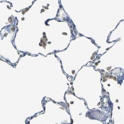
- Experimental details
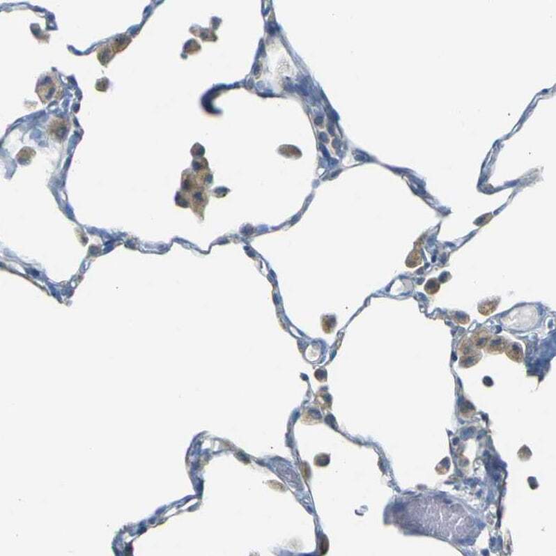
- Immunohistochemical staining of PARD6B in human lung using PARD6B Polyclonal Antibody (Product # PA5-53050) shows moderate cytoplasmic positivity in macrophages.
- Submitted by
- Invitrogen Antibodies (provider)
- Main image

- Experimental details
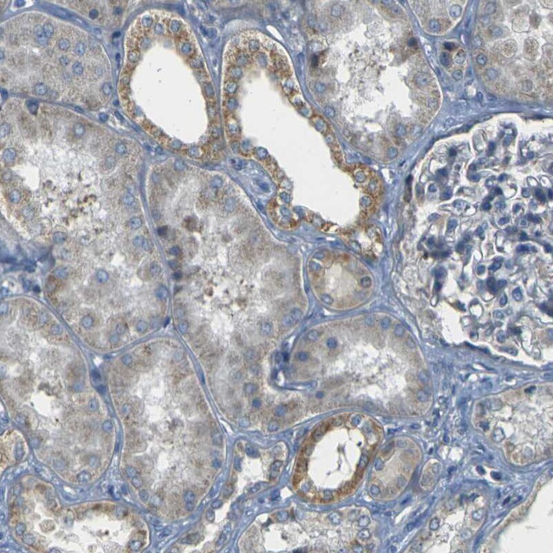
- Immunohistochemical staining of PARD6B in human kidney using PARD6B Polyclonal Antibody (Product # PA5-53050) shows weak to moderate cytoplasmic positivity in cells in tubules.
- Submitted by
- Invitrogen Antibodies (provider)
- Main image
- Experimental details
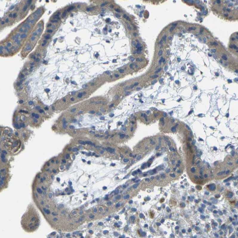
- Immunohistochemical staining of PARD6B in human placenta using PARD6B Polyclonal Antibody (Product # PA5-53050) shows weak cytoplasmic positivity in trophoblastic cells.
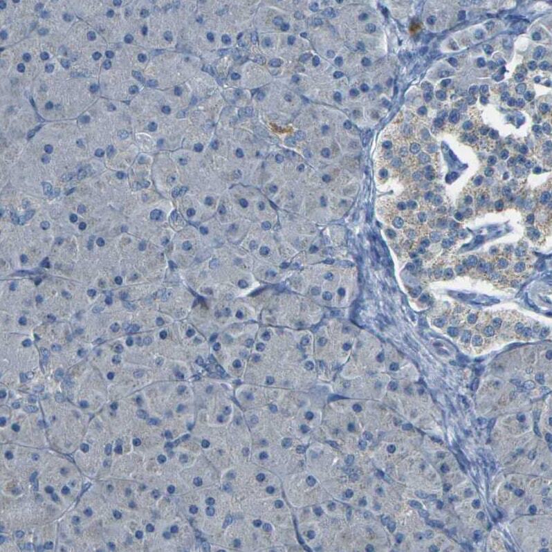
- Submitted by
- Invitrogen Antibodies (provider)
- Main image
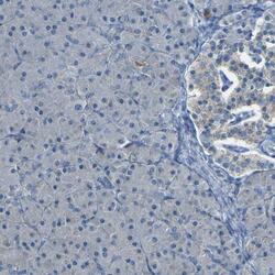
- Experimental details
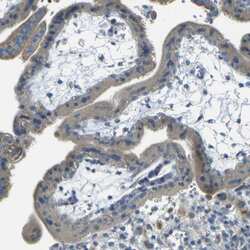
- Immunohistochemical staining of PARD6B in human pancreas using PARD6B Polyclonal Antibody (Product # PA5-53050) shows very weak positivity in exocrine glandular cells.