Explore
 Validate
 Learn
Learn Western blot
 Immunocytochemistry
Antibody data
- Antibody Data
- Antigen structure
- References [0]
- Comments [0]
- Validations
- Immunocytochemistry [5]
- Immunohistochemistry [1]
Submit
Validation data
Reference
Comment
Report error
- Product number
- PA5-34916 - Provider product page

- Provider
- Invitrogen Antibodies
- Product name
- SLU7 Polyclonal Antibody
- Antibody type
- Polyclonal
- Antigen
- Synthetic peptide
- Description
- Recommended positive controls: 293T, A431, HeLa, HepG2. Predicted reactivity: Mouse (100%), Rat (100%), Rhesus Monkey (100%), Chimpanzee (100%), Bovine (100%). Store product as a concentrated solution. Centrifuge briefly prior to opening the vial.
- Reactivity
- Human
- Host
- Rabbit
- Isotype
- IgG
- Vial size
- 100 μL
- Concentration
- 1 mg/mL
- Storage
- Store at 4°C short term. For long term storage, store at -20°C, avoiding freeze/thaw cycles.
No comments: Submit comment
Supportive validation
- Submitted by
- Invitrogen Antibodies (provider)
- Main image
- Experimental details
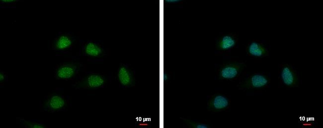
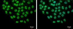
- Immunofluorescent analysis of Slu7 showing staining in the nucleus of HeLa cells. HeLa cells were fixed in 4% paraformaldehyde at RT for 15 min and stained using a Slu7 polyclonal antibody (Product # PA5-34916) diluted at 1:500. Blue: Hoechst 33342 staining. Scale bar = 10µm.
- Submitted by
- Invitrogen Antibodies (provider)
- Main image
- Experimental details
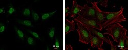
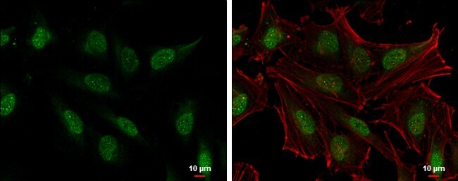
- Immunocytochemistry-Immunofluorescence analysis of SLU7 was performed in HeLa cells fixed in 4% paraformaldehyde at RT for 15 min. Green: SLU7 Polyclonal Antibody (Product # PA5-34916) diluted at 1:500. Red: phalloidin, a cytoskeleton marker. Blue: Hoechst 33342 staining. Scale bar = 10 µm.
- Submitted by
- Invitrogen Antibodies (provider)
- Main image

- Experimental details
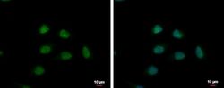
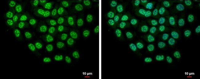
- Slu7 antibody detects Slu7 protein at nucleus by immunofluorescent analysis. Sample: HCT116 cells were fixed in 4% paraformaldehyde at RT for 15 min. Green: Slu7 protein stained by Slu7 antibody (Product # PA5-34916) diluted at 1:500. Blue: Hoechst 33342 staining. Scale bar = 10 µm.
- Submitted by
- Invitrogen Antibodies (provider)
- Main image
- Experimental details
- Slu7 antibody detects Slu7 protein at nucleus by immunofluorescent analysis. Sample: HCT116 cells were fixed in 4% paraformaldehyde at RT for 15 min. Green: Slu7 protein stained by Slu7 antibody (Product # PA5-34916) diluted at 1:500. Blue: Hoechst 33342 staining. Scale bar = 10 µm.

- Submitted by
- Invitrogen Antibodies (provider)
- Main image

- Experimental details
- Immunocytochemistry-Immunofluorescence analysis of SLU7 was performed in HeLa cells fixed in 4% paraformaldehyde at RT for 15 min. Green: SLU7 Polyclonal Antibody (Product # PA5-34916) diluted at 1:500. Red: phalloidin, a cytoskeleton marker. Blue: Hoechst 33342 staining. Scale bar = 10 µm.

Supportive validation
- Submitted by
- Invitrogen Antibodies (provider)
- Main image
- Experimental details
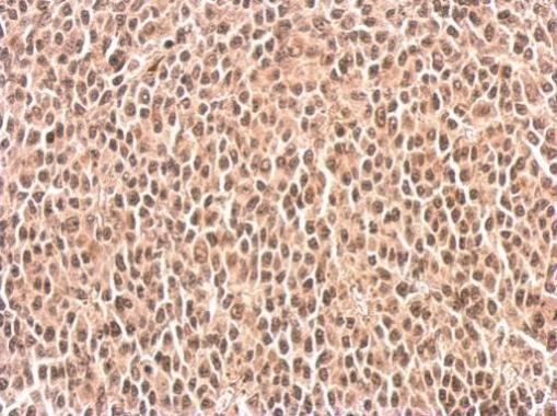
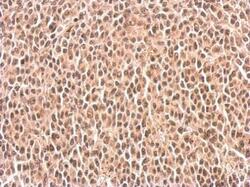
- Slu7 antibody detects SLU7 protein at nucleus on BT483 xenograft by immunohistochemical analysis. Sample: Paraffin-embedded BT483 xenograft. Slu7 antibody (Product # PA5-34916) dilution: 1:500. Antigen Retrieval: EDTA based buffer, pH 8.0, 15 min.