Explore
 Validate
 Learn
Learn Flow cytometry
Antibody data
- Antibody Data
- Antigen structure
- References [0]
- Comments [0]
- Validations
- Flow cytometry [3]
Submit
Validation data
Reference
Comment
Report error
- Product number
- Ab108247 - Provider product page

- Provider
- Aladdin Scientific
- Product name
- HLA-DR Mouse mAb
- Antibody type
- Monoclonal
- Description
- Mouse anti Human HLA-DR Antibody, Monoclonal (L243), could be used for Flow and so on.Application:Flow: 0.06 _g/100w cells Protein FunctionBinds peptides derived from antigens that access the endocytic route of antigen presenting cells (APC) and presents them on the cell surface for recognition by the CD4 T-cells. The peptide binding cleft accommodates peptides of 10-30 residues. The peptides presented by MHC class II molecules are generated mostly by degradation of proteins that access the endocytic route, where they are processed by lysosomal proteases and other hydrolases. Exogenous antigens that have been endocytosed by the APC are thus readily available for presentation via MHC II molecules, and for this reason this antigen presentation pathway is usually referred to as exogenous. As membrane proteins on their way to degradation in lysosomes as part of their normal turn-over are also contained in the endosomal/lysosomal compartments, exogenous antigens must compete with those derived from endogenous components. Autophagy is also a source of endogenous peptides, autophagosomes constitutively fuse with MHC class II loading compartments. In addition to APCs, other cells of the gastrointestinal tract, such as epithelial cells, express MHC class II molecules and CD74 and act as APCs, which is an unusual trait of the GI tract. To produce a MHC class II molecule that presents an antigen, three MHC class II molecules (heterodimers of an alpha and a beta chain) associate with a CD74 trimer in the ER to form an heterononamer. Soon after the entry of this complex into the endosomal/lysosomal system where antigen processing occurs, CD74 undergoes a sequential degradation by various proteases, including CTSS and CTSL, leaving a small fragment termed CLIP (class-II-associated invariant chain peptide). The removal of CLIP is facilitated by HLA-DM via direct binding to the alpha-beta-CLIP complex so that CLIP is released. HLA-DM stabilizes MHC class II molecules until primary high affinity antigenic peptides are bound. The MHC II molecule bound to a peptide is then transported to the cell membrane surface. In B-cells, the interaction between HLA-DM and MHC class II molecules is regulated by HLA-DO. Primary dendritic cells (DCs) also to express HLA-DO. Lysosomal miroenvironment has been implicated in the regulation of antigen loading into MHC II molecules, increased acidification produces increased proteolysis and efficient peptide loading.
- Reactivity
- Human
- Host
- Mouse
- Conjugate
- Unconjugated
- Antibody clone number
- L243
- Vial size
- 100_g,10_g,1mg,50_g
- Concentration
- Lot by Lot
- Storage
- Shipped at 4¡C. Store at 4¡C long term (12 months). Upon delivery aliquot.
No comments: Submit comment
Supportive validation
- Submitted by
- Aladdin Scientific (provider)
- Main image

- Experimental details
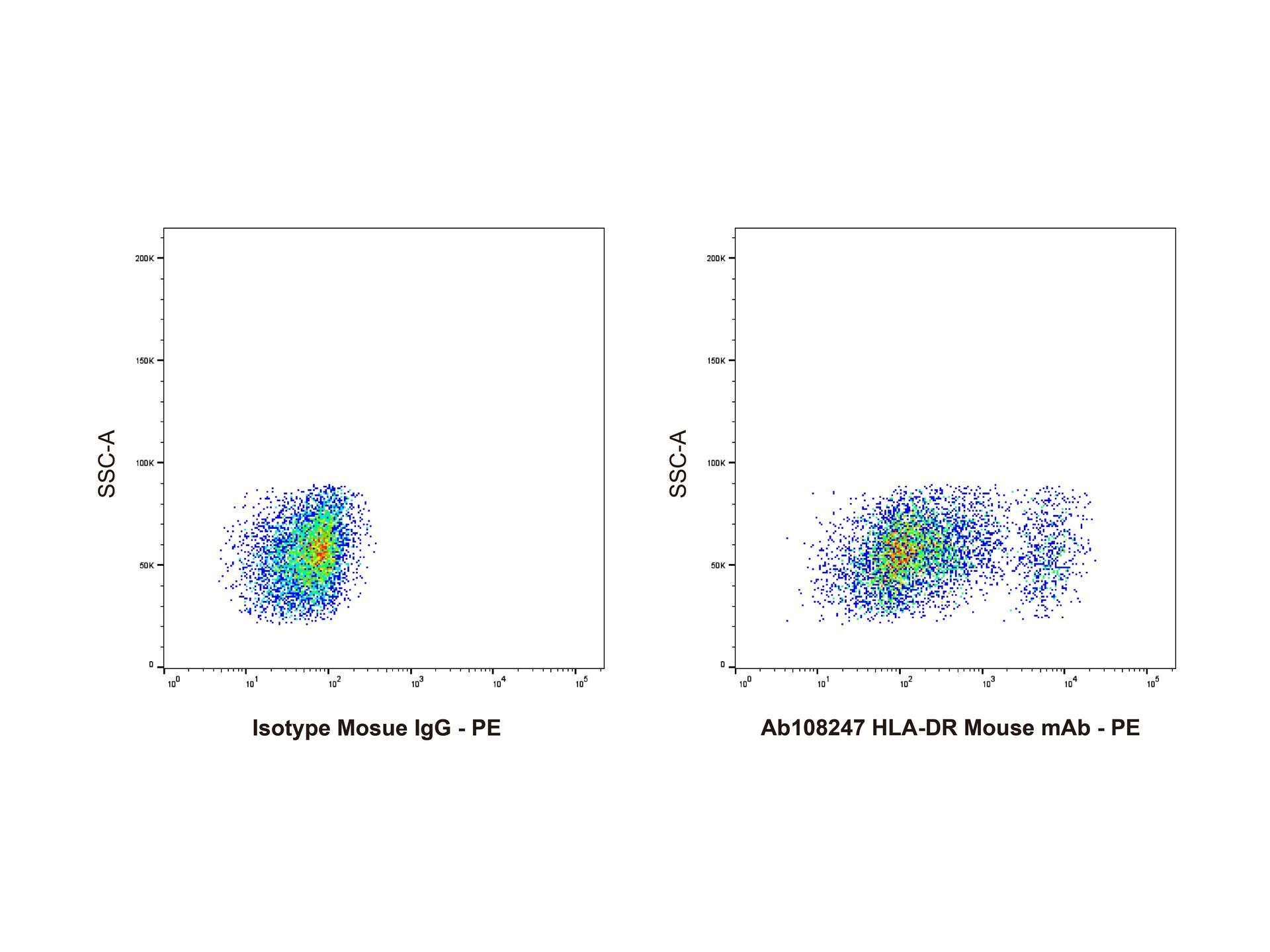
- HLA-DR Mouse mAb (Ab108247) - Flow Cytometry Flow Cytometry analysis of human peripheral blood lymphocytes labelling HLA-DR with HLA-DR Mouse mAb (Ab108247) conjugated with biotin (right panel) at 0.1 μg/mL for 1 hour at 4°C, compared with mouse IgG (Ab170221) conjugated with biotin - Isotype Control (left panel) and detected with SA-PE (rp156250).
- Submitted by
- Aladdin Scientific (provider)
- Main image
- Experimental details
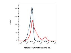
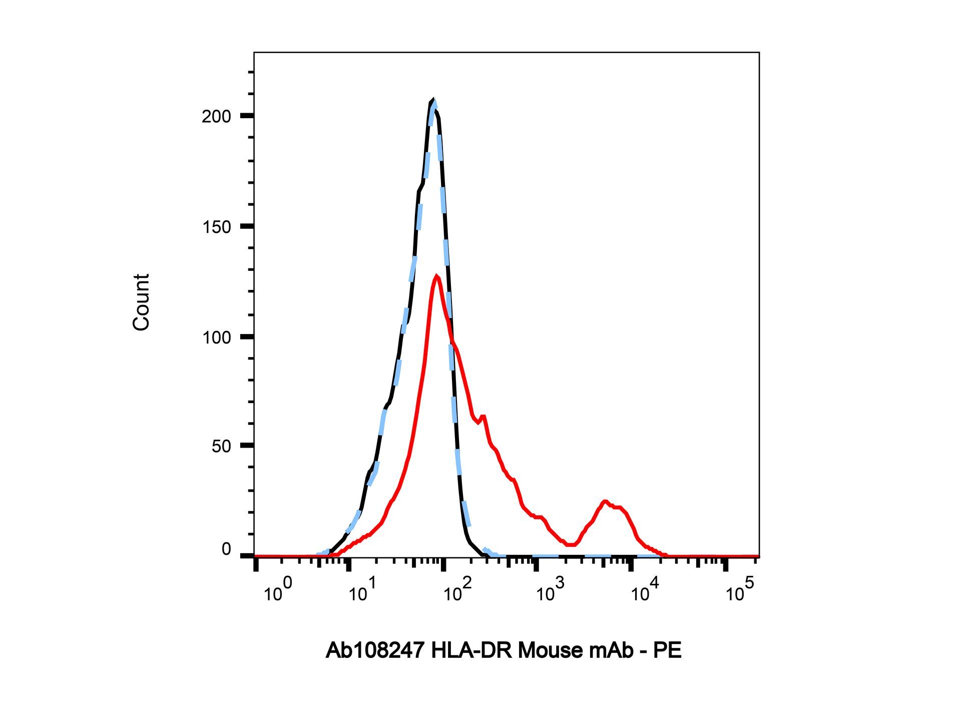
- HLA-DR Mouse mAb (Ab108247) - Flow Cytometry Flow Cytometry analysis of human peripheral blood lymphocytes labelling HLA-DR (red) with HLA-DR Mouse mAb (Ab108247) conjugated with biotin at 0.1 μg/mL for 1 hour at 4°C, and detected with SA-PE (rp156250). Blue - Isotype control, mouse IgG (Ab170221) conjugated with biotin. Black - Unlabelled control, cells without incubation with primary antibody.
- Submitted by
- Aladdin Scientific (provider)
- Main image
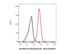
- Experimental details
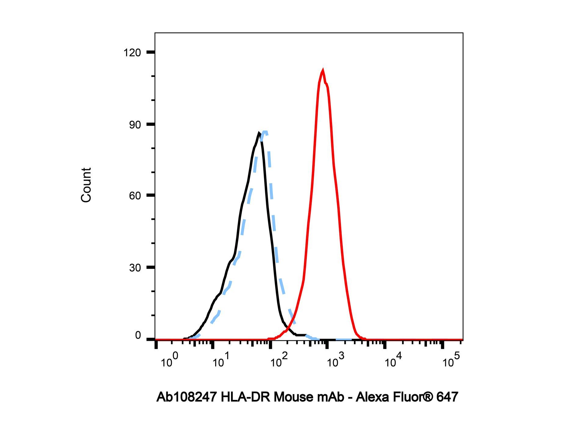
- HLA-DR Mouse mAb (Ab108247) - Flow Cytometry Flow Cytometry analysis of Raji cells labelling HLA-DR (red) with HLA-DR Mouse mAb (Ab108247) at 0.1 μg/mL for 1 hour at 4°C. AffiniPure Goat anti-Mouse IgG, Fcγ fragment specific (Alexa Fluor® 647) at a dilution of 1/1000 was used as the secondary antibody. Blue - Isotype control, mouse IgG (Ab170221). Black - Unlabelled control, cells without incubation with primary antibody.