Explore
 Validate
 Learn
Learn Western blot
 Immunohistochemistry
Antibody data
- Antibody Data
- Antigen structure
- References [1]
- Comments [0]
- Validations
- Western blot [1]
- Other assay [1]
Submit
Validation data
Reference
Comment
Report error
- Product number
- PA5-77404 - Provider product page

- Provider
- Invitrogen Antibodies
- Product name
- GABRG2 (extracellular) Polyclonal Antibody
- Antibody type
- Polyclonal
- Antigen
- Synthetic peptide
- Description
- For reconstitution, we recommend adding 100 µL distilled water to a final antibody concentration of about 1 mg/mL. To use this carrier-free antibody for conjugation experiments, we strongly recommend performing another round of desalting. (Zeba Spin Desalting Columns, 7KMWCO, 0.5 mL, Product # 89882)
- Reactivity
- Human, Mouse, Rat
- Host
- Rabbit
- Isotype
- IgG
- Vial size
- 50 µL
- Concentration
- 0.8 mg/mL
- Storage
- -20°C
Submitted references Neocortex- and hippocampus-specific deletion of Gabrg2 causes temperature-dependent seizures in mice.
Li X, Guo S, Xu S, Chen Z, Wang L, Ding J, Huo J, Xiao L, He Z, Jin Z, Wang F, Sun T
Cell death & disease 2021 May 28;12(6):553
Cell death & disease 2021 May 28;12(6):553
No comments: Submit comment
Supportive validation
- Submitted by
- Invitrogen Antibodies (provider)
- Main image

- Experimental details
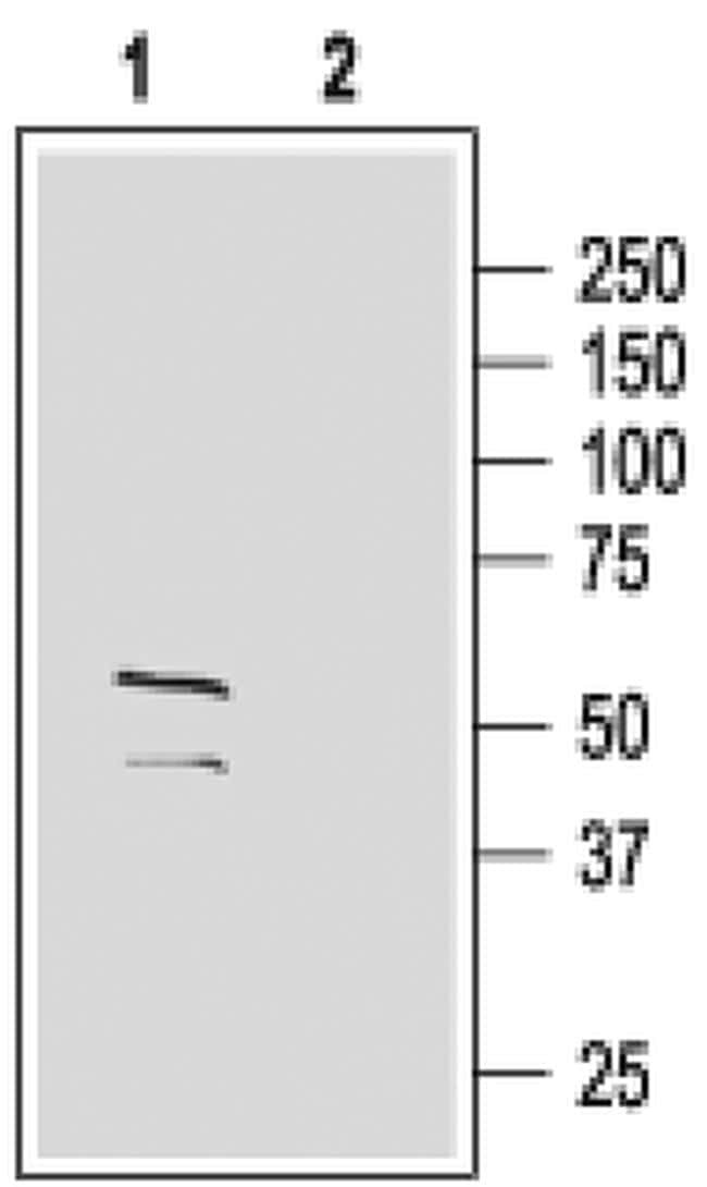
- Western blot analysis of rat brain membranes with GABRG2 (extracellular) polyclonal antibody (Product # PA5-77404) using a dilution of 1:200.
Supportive validation
- Submitted by
- Invitrogen Antibodies (provider)
- Main image
- Experimental details
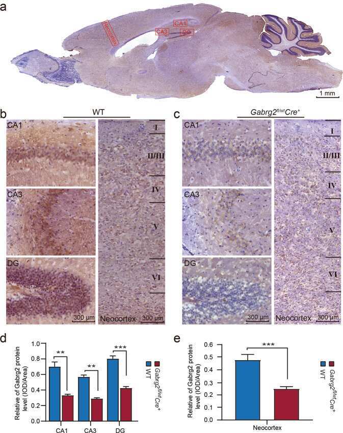
- Fig. 4 Immunohistochemistry of neurons in the mouse brain. a The immunohistochemistry of the Gabrg2 fl/wt Cre + mice brain was performed. The CA1, CA3, and DG, and neocortex regions were selected in a sagittal section. This mouse brain section was immunostained with an anti-Gabrg2 antibody, showing brown stain. Scale bar: 1 mm. b Representative immunohistochemical staining of the hippocampus and cerebral cortex of WT mice using the antibody against Gabrg2, where most of neurons in CA1, CA3 and DG, and the neocortex regions were stained. Compared with Gabrg2 fl/wt Cre + mice, neurons in the hippocampus and neocortex organised in groups in the WT are neat, relatively close, and have a lot of synapses. Scale bar: 300 um. c Representative immunohistochemistry images showing the Gabrg2 proteins in the CA1, CA3, and DG, and neocortex regions of Gabrg2 fl/wt Cre + mice. The density of stained neurons was significantly lower than that of the control group. Scale bar: 300 um. d Histograms of the Gabrg2 protein expression in the hippocampus (CA1, CA3 and DG) quantified from immunohistochemistry analysis in WT ( n = 4) and Gabrg2 fl/wt Cre + mice ( n = 6). e The relative Gabrg2 protein level in the neocortex of WT and Gabrg2 fl/wt Cre + mice ( n = 4 for WT and n = 6 for KO mice). Data shown are mean +- standard error of mean. ** P < 0.01 and *** P < 0.001 vs WT, t -test (two-tailed).