Explore
 Validate
 Learn
Learn Western blot
Antibody data
- Antibody Data
- Antigen structure
- References [3]
- Comments [0]
- Validations
- Western blot [4]
- Immunohistochemistry [1]
Submit
Validation data
Reference
Comment
Report error
- Product number
- GTX113574 - Provider product page

- Provider
- GeneTex
- Proper citation
- GeneTex Cat#GTX113574, RRID:AB_10620764
- Product name
- VEGFC antibody
- Antibody type
- Polyclonal
- Reactivity
- Human, Mouse, Rat
- Host
- Rabbit
Submitted references High Glucose Induces VEGF-C Expression via the LPA1/3-Akt-ROS-LEDGF Signaling Axis in Human Prostate Cancer PC-3 Cells.
LPA1/3 signaling mediates tumor lymphangiogenesis through promoting CRT expression in prostate cancer.
Interleukin-6 Induces Vascular Endothelial Growth Factor-C Expression via Src-FAK-STAT3 Signaling in Lymphatic Endothelial Cells.
Huang YL, Lin YC, Lin CC, Chen WM, Chen BPC, Lee H
Cellular physiology and biochemistry : international journal of experimental cellular physiology, biochemistry, and pharmacology 2018;50(2):597-611
Cellular physiology and biochemistry : international journal of experimental cellular physiology, biochemistry, and pharmacology 2018;50(2):597-611
LPA1/3 signaling mediates tumor lymphangiogenesis through promoting CRT expression in prostate cancer.
Lin YC, Chen CC, Chen WM, Lu KY, Shen TL, Jou YC, Shen CH, Ohbayashi N, Kanaho Y, Huang YL, Lee H
Biochimica et biophysica acta. Molecular and cell biology of lipids 2018 Oct;1863(10):1305-1315
Biochimica et biophysica acta. Molecular and cell biology of lipids 2018 Oct;1863(10):1305-1315
Interleukin-6 Induces Vascular Endothelial Growth Factor-C Expression via Src-FAK-STAT3 Signaling in Lymphatic Endothelial Cells.
Huang YH, Yang HY, Huang SW, Ou G, Hsu YF, Hsu MJ
PloS one 2016;11(7):e0158839
PloS one 2016;11(7):e0158839
No comments: Submit comment
Supportive validation
- Submitted by
- GeneTex (provider)
- Main image
- Experimental details
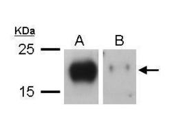
- Sample (20 ?g of whole cell lysate) A: TL1 whole cell lysate B: TL1 knock down VEGF-C whole cell lysate 12% SDS PAGE GTX113574 diluted at 1:1000 The HRP-conjugated anti-rabbit IgG antibody (GTX213110-01) was used to detect the primary antibody.

- Submitted by
- GeneTex (provider)
- Main image
- Experimental details
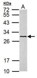
- VEGFC antibody detects VEGFC protein by western blot analysis. A. 50 ?g mouse lung lysate/extract 12% SDS-PAGE VEGFC antibody (GTX113574) dilution: 1:1000 The HRP-conjugated anti-rabbit IgG antibody (GTX213110-01) was used to detect the primary antibody.
- Submitted by
- GeneTex (provider)
- Main image
- Experimental details
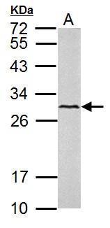
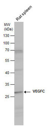
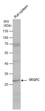
- VEGFC antibody detects VEGFC protein by western blot analysis. Rat tissue extracts (50 ?g) was separated by 10% SDS-PAGE, and the membrane was blotted with VEGFC antibody (GTX113574) diluted at 1:1000. The HRP-conjugated anti-rabbit IgG antibody (GTX213110-01) was used to detect the primary antibody.
- Submitted by
- GeneTex (provider)
- Main image
- Experimental details
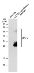
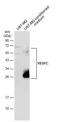
- U87-MG whole cell extract and conditioned medium (30 ?g) were separated by 12% SDS-PAGE, and the membrane was blotted with VEGFC antibody (GTX113574) diluted at 1:500. The HRP-conjugated anti-rabbit IgG antibody (GTX213110-01) was used to detect the primary antibody.
Supportive validation
- Submitted by
- GeneTex (provider)
- Main image
- Experimental details
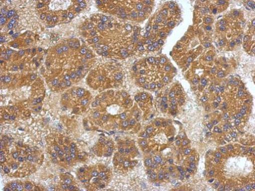
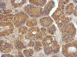
- Immunohistochemical analysis of paraffin-embedded human hepatoma, using VEGFC(GTX113574) antibody at 1:500 dilution.