 Explore
 Validate
 Learn
Learn Western blot
Antibody data
- Antibody Data
- Antigen structure
- References [11]
- Comments [0]
- Validations
- Western blot [1]
- Immunohistochemistry [2]
Submit
Validation data
Reference
Comment
Report error
- Product number
- AF752 - Provider product page

- Provider
- R&D Systems
- Product name
- Human VEGF-C Antibody
- Antibody type
- Polyclonal
- Description
- Antigen Affinity-purified. Detects human VEGF-C in direct ELISAs and Western blots. In direct ELISAs, less than 1% cross-reactivity with recombinant human (rh) VEGF-D and rhVEGF-A is observed.
- Reactivity
- Human
- Host
- Goat
- Conjugate
- Unconjugated
- Isotype
- IgG
- Vial size
- 100 ug
- Concentration
- LYOPH
- Storage
- Use a manual defrost freezer and avoid repeated freeze-thaw cycles. 12 months from date of receipt, -20 to -70 °C as supplied. 1 month, 2 to 8 °C under sterile conditions after reconstitution. 6 months, -20 to -70 °C under sterile conditions after reconstitution.
Submitted references The interplay of matrix metalloproteinase-8, transforming growth factor-β1 and vascular endothelial growth factor-C cooperatively contributes to the aggressiveness of oral tongue squamous cell carcinoma.
Synaptonemal complex protein 3 is associated with lymphangiogenesis in non-small cell lung cancer patients with lymph node metastasis.
Efficient activation of the lymphangiogenic growth factor VEGF-C requires the C-terminal domain of VEGF-C and the N-terminal domain of CCBE1.
A phase Ib/II translational study of sunitinib with neoadjuvant radiotherapy in soft-tissue sarcoma.
Lysophosphatidic acid enhances vascular endothelial growth factor-C expression in human prostate cancer PC-3 cells.
Dilated thin-walled blood and lymphatic vessels in human endometrium: a potential role for VEGF-D in progestin-induced break-through bleeding.
Autocrine loop between vascular endothelial growth factor (VEGF)-C and VEGF receptor-3 positively regulates tumor-associated lymphangiogenesis in oral squamoid cancer cells.
Lymphatic reprogramming of microvascular endothelial cells by CEA-related cell adhesion molecule-1 via interaction with VEGFR-3 and Prox1.
VEGF-C is a trophic factor for neural progenitors in the vertebrate embryonic brain.
Cyclooxygenase-2 induces EP1- and HER-2/Neu-dependent vascular endothelial growth factor-C up-regulation: a novel mechanism of lymphangiogenesis in lung adenocarcinoma.
Cyclooxygenase-2 induces EP1- and HER-2/Neu-dependent vascular endothelial growth factor-C up-regulation: a novel mechanism of lymphangiogenesis in lung adenocarcinoma.
Åström P, Juurikka K, Hadler-Olsen ES, Svineng G, Cervigne NK, Coletta RD, Risteli J, Kauppila JH, Skarp S, Kuttner S, Oteiza A, Sutinen M, Salo T
British journal of cancer 2017 Sep 26;117(7):1007-1016
British journal of cancer 2017 Sep 26;117(7):1007-1016
Synaptonemal complex protein 3 is associated with lymphangiogenesis in non-small cell lung cancer patients with lymph node metastasis.
Kitano H, Chung JY, Noh KH, Lee YH, Kim TW, Lee SH, Eo SH, Cho HJ, Choi CH, Inoue S, Hanaoka J, Fukuoka J, Hewitt SM
Journal of translational medicine 2017 Jun 17;15(1):138
Journal of translational medicine 2017 Jun 17;15(1):138
Efficient activation of the lymphangiogenic growth factor VEGF-C requires the C-terminal domain of VEGF-C and the N-terminal domain of CCBE1.
Jha SK, Rauniyar K, Karpanen T, Leppänen VM, Brouillard P, Vikkula M, Alitalo K, Jeltsch M
Scientific reports 2017 Jul 7;7(1):4916
Scientific reports 2017 Jul 7;7(1):4916
A phase Ib/II translational study of sunitinib with neoadjuvant radiotherapy in soft-tissue sarcoma.
Lewin J, Khamly KK, Young RJ, Mitchell C, Hicks RJ, Toner GC, Ngan SY, Chander S, Powell GJ, Herschtal A, Te Marvelde L, Desai J, Choong PF, Stacker SA, Achen MG, Ferris N, Fox S, Slavin J, Thomas DM
British journal of cancer 2014 Dec 9;111(12):2254-61
British journal of cancer 2014 Dec 9;111(12):2254-61
Lysophosphatidic acid enhances vascular endothelial growth factor-C expression in human prostate cancer PC-3 cells.
Lin CE, Chen SU, Lin CC, Chang CH, Lin YC, Tai YL, Shen TL, Lee H
PloS one 2012;7(7):e41096
PloS one 2012;7(7):e41096
Dilated thin-walled blood and lymphatic vessels in human endometrium: a potential role for VEGF-D in progestin-induced break-through bleeding.
Donoghue JF, McGavigan CJ, Lederman FL, Cann LM, Fu L, Dimitriadis E, Girling JE, Rogers PA
PloS one 2012;7(2):e30916
PloS one 2012;7(2):e30916
Autocrine loop between vascular endothelial growth factor (VEGF)-C and VEGF receptor-3 positively regulates tumor-associated lymphangiogenesis in oral squamoid cancer cells.
Matsuura M, Onimaru M, Yonemitsu Y, Suzuki H, Nakano T, Ishibashi H, Shirasuna K, Sueishi K
The American journal of pathology 2009 Oct;175(4):1709-21
The American journal of pathology 2009 Oct;175(4):1709-21
Lymphatic reprogramming of microvascular endothelial cells by CEA-related cell adhesion molecule-1 via interaction with VEGFR-3 and Prox1.
Kilic N, Oliveira-Ferrer L, Neshat-Vahid S, Irmak S, Obst-Pernberg K, Wurmbach JH, Loges S, Kilic E, Weil J, Lauke H, Tilki D, Singer BB, Ergün S
Blood 2007 Dec 15;110(13):4223-33
Blood 2007 Dec 15;110(13):4223-33
VEGF-C is a trophic factor for neural progenitors in the vertebrate embryonic brain.
Le Bras B, Barallobre MJ, Homman-Ludiye J, Ny A, Wyns S, Tammela T, Haiko P, Karkkainen MJ, Yuan L, Muriel MP, Chatzopoulou E, Bréant C, Zalc B, Carmeliet P, Alitalo K, Eichmann A, Thomas JL
Nature neuroscience 2006 Mar;9(3):340-8
Nature neuroscience 2006 Mar;9(3):340-8
Cyclooxygenase-2 induces EP1- and HER-2/Neu-dependent vascular endothelial growth factor-C up-regulation: a novel mechanism of lymphangiogenesis in lung adenocarcinoma.
Su JL, Shih JY, Yen ML, Jeng YM, Chang CC, Hsieh CY, Wei LH, Yang PC, Kuo ML
Cancer research 2004 Jan 15;64(2):554-64
Cancer research 2004 Jan 15;64(2):554-64
No comments: Submit comment
Supportive validation
- Submitted by
- R&D Systems (provider)
- Main image
- Experimental details
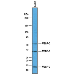
- Detection of Human VEGF-C by Western Blot. Western blot shows lysates of K562 human chronic myelogenous leukemia cell line. PVDF membrane was probed with 1 µg/mL of Goat Anti-Human VEGF-C Antigen Affinity-purified Polyclonal Antibody (Catalog # AF752) followed by HRP-conjugated Anti-Goat IgG Secondary Antibody (Catalog # HAF017). Specific bands were detected for VEGF-C at approximately 52 kDa, 34 kDa, and 13 kDa (as indicated). This experiment was conducted under reducing conditions and using Immunoblot Buffer Group 1.

Supportive validation
- Submitted by
- R&D Systems (provider)
- Main image

- Experimental details
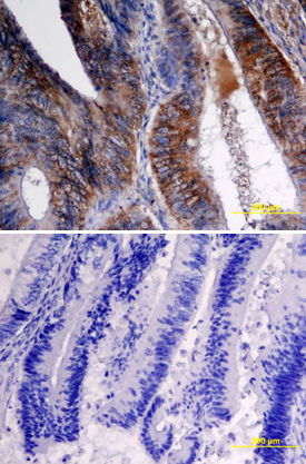
- VEGF-C in Human Colon Cancer Tissue. VEGF-C was detected in immersion fixed paraffin-embedded sections of human colon cancer tissue using Goat Anti-Human VEGF-C Antigen Affinity-purified Polyclonal Antibody (Catalog # AF752) at 15 µg/mL overnight at 4 °C. Tissue was stained using the Anti-Goat HRP-DAB Cell & Tissue Staining Kit (brown; Catalog # CTS008) and counterstained with hematoxylin (blue). Specific labeling was localized to epithelial cells in crypts of the colon mucusa (longitudinal section of crypts). Lower panel shows a lack of labeling if primary antibodies are omitted and tissue is stained only with secondary antibody followed by incubation with detection reagents. View our protocol for Chromogenic IHC Staining of Paraffin-embedded Tissue Sections.
- Submitted by
- R&D Systems (provider)
- Main image
- Experimental details
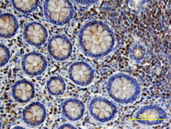
- VEGF-C in Human Colon Cancer Tissue. VEGF-C was detected in immersion fixed paraffin-embedded sections of human colon cancer tissue using Goat Anti-Human VEGF-C Antigen Affinity-purified Polyclonal Antibody (Catalog # AF752) at 10 µg/mL overnight at 4 °C. Tissue was stained using the Anti-Goat HRP-DAB Cell & Tissue Staining Kit (brown; Catalog # CTS008) and counterstained with hematoxylin (blue). Specific labeling was localized to stromal cells surrounding crypts in the colon mucosa (cross section across crypts). View our protocol for Chromogenic IHC Staining of Paraffin-embedded Tissue Sections.
