 Explore
 Validate
 Learn
Learn Western blot
Antibody data
- Antibody Data
- Antigen structure
- References [1]
- Comments [0]
- Validations
- Western blot [1]
- Immunohistochemistry [1]
Submit
Validation data
Reference
Comment
Report error
- Product number
- MAB7557 - Provider product page

- Provider
- R&D Systems
- Product name
- Human CIB1 Antibody
- Antibody type
- Monoclonal
- Description
- Protein A or G purified from hybridoma culture supernatant. Detects human CIB1 in direct ELISAs and Western blots.
- Reactivity
- Human
- Host
- Mouse
- Conjugate
- Unconjugated
- Antigen sequence
E9Q3T6- Isotype
- IgG
- Antibody clone number
- 791119
- Vial size
- 100 ug
- Concentration
- LYOPH
- Storage
- Use a manual defrost freezer and avoid repeated freeze-thaw cycles. 12 months from date of receipt, -20 to -70 °C as supplied. 1 month, 2 to 8 °C under sterile conditions after reconstitution. 6 months, -20 to -70 °C under sterile conditions after reconstitution.
Submitted references CIB1 contributes to oncogenic signalling by Ras via modulating the subcellular localisation of sphingosine kinase 1.
Zhu W, Gliddon BL, Jarman KE, Moretti PAB, Tin T, Parise LV, Woodcock JM, Powell JA, Ruszkiewicz A, Pitman MR, Pitson SM
Oncogene 2017 May 4;36(18):2619-2627
Oncogene 2017 May 4;36(18):2619-2627
No comments: Submit comment
Supportive validation
- Submitted by
- R&D Systems (provider)
- Main image
- Experimental details
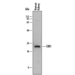
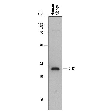
- Detection of Human CIB1 by Western Blot. Western blot shows lysates of human kidney tissue. PVDF membrane was probed with 2 µg/mL of Mouse Anti-Human CIB1 Monoclonal Antibody (Catalog # MAB7557) followed by HRP-conjugated Anti-Mouse IgG Secondary Antibody (Catalog # HAF018). A specific band was detected for CIB1 at approximately 22 kDa (as indicated). This experiment was conducted under reducing conditions and using Immunoblot Buffer Group 1.
Supportive validation
- Submitted by
- R&D Systems (provider)
- Main image
- Experimental details
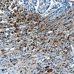
- CIB1 in Human Breast Cancer Tissue. CIB1 was detected in immersion fixed paraffin-embedded sections of human breast cancer tissue using Mouse Anti-Human CIB1 Monoclonal Antibody (Catalog # MAB7557) at 15 µg/mL overnight at 4 °C. Before incubation with the primary antibody, tissue was subjected to heat-induced epitope retrieval using Antigen Retrieval Reagent-Basic (Catalog # CTS013). Tissue was stained using the Anti-Mouse HRP-DAB Cell & Tissue Staining Kit (brown; Catalog # CTS002) and counterstained with hematoxylin (blue). Specific staining was localized to cytoplasm of cancer cells. View our protocol for Chromogenic IHC Staining of Paraffin-embedded Tissue Sections.
