Explore
 Validate
 Learn
Learn Western blot
 Immunocytochemistry
Antibody data
- Antibody Data
- Antigen structure
- References [2]
- Comments [0]
- Validations
- Immunocytochemistry [3]
- Immunohistochemistry [1]
- Other assay [2]
Submit
Validation data
Reference
Comment
Report error
- Product number
- PA5-21671 - Provider product page

- Provider
- Invitrogen Antibodies
- Product name
- PP1 gamma Polyclonal Antibody
- Antibody type
- Polyclonal
- Antigen
- Recombinant full-length protein
- Description
- Recommended positive controls: 293T, A431, HeLa, HepG2, Molt-4, Raji, NIH-3T3. Predicted reactivity: Mouse (100%), Rat (100%), Xenopus laevis (100%), Dog (99%), Pig (100%), Chicken (100%), Rhesus Monkey (100%), Bovine (100%). Store product as a concentrated solution. Centrifuge briefly prior to opening the vial.
- Reactivity
- Human, Mouse
- Host
- Rabbit
- Isotype
- IgG
- Vial size
- 100 μL
- Concentration
- 0.79 mg/mL
- Storage
- Store at 4°C short term. For long term storage, store at -20°C, avoiding freeze/thaw cycles.
Submitted references Ultrastructural Localization and Molecular Associations of HCV Capsid Protein in Jurkat T Cells.
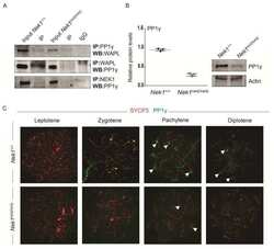
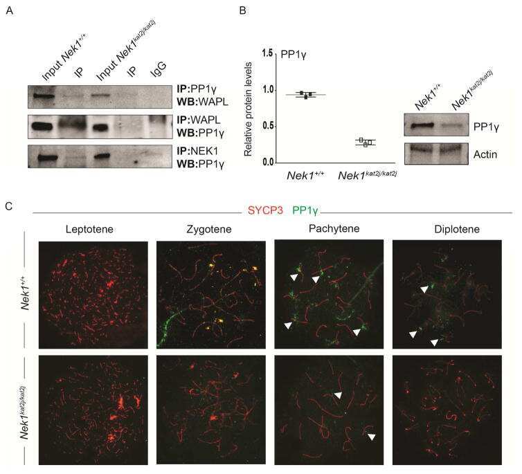
Cohesin Removal along the Chromosome Arms during the First Meiotic Division Depends on a NEK1-PP1γ-WAPL Axis in the Mouse.
Fernández-Ponce C, Durán-Ruiz MC, Narbona-Sánchez I, Muñoz-Miranda JP, Arbulo-Echevarria MM, Serna-Sanz A, Baumann C, Litrán R, Aguado E, Bloch W, García-Cozar F
Frontiers in microbiology 2017;8:2595
Frontiers in microbiology 2017;8:2595
Cohesin Removal along the Chromosome Arms during the First Meiotic Division Depends on a NEK1-PP1γ-WAPL Axis in the Mouse.
Brieño-Enríquez MA, Moak SL, Toledo M, Filter JJ, Gray S, Barbero JL, Cohen PE, Holloway JK
Cell reports 2016 Oct 18;17(4):977-986
Cell reports 2016 Oct 18;17(4):977-986
No comments: Submit comment
Supportive validation
- Submitted by
- Invitrogen Antibodies (provider)
- Main image
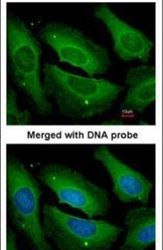
- Experimental details
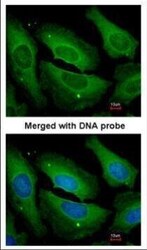
- Immunofluorescent analysis of PP1 gamma in paraformaldehyde-fixed HeLa cells using a PP1 gamma polyclonal antibody (Product # PA5-21671) at a 1:200 dilution.
- Submitted by
- Invitrogen Antibodies (provider)
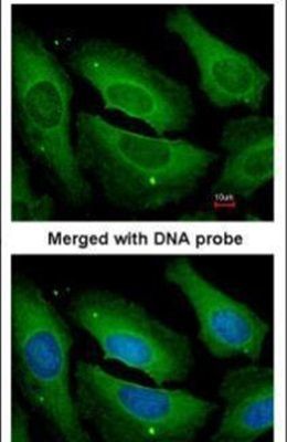
- Main image

- Experimental details
- Immunofluorescence analysis of paraformaldehyde-fixed HeLa, using PP1 gamma antibody (Product # PA5-21671) at 1:200 dilution.
- Submitted by
- Invitrogen Antibodies (provider)
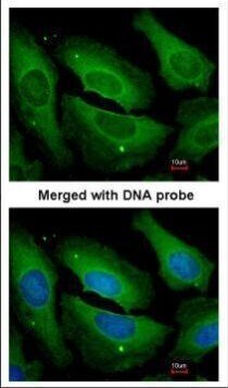
- Main image
- Experimental details
- Immunofluorescence analysis of paraformaldehyde-fixed HeLa, using PP1 gamma antibody (Product # PA5-21671) at 1:200 dilution.

Supportive validation
- Submitted by
- Invitrogen Antibodies (provider)
- Main image

- Experimental details
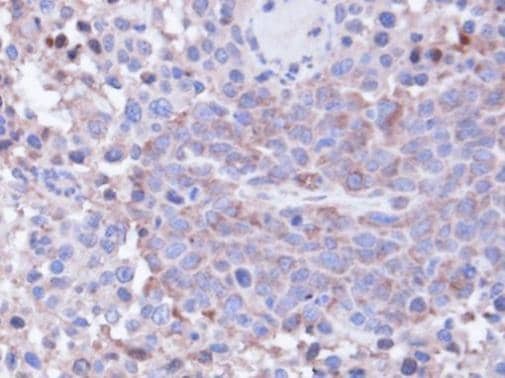
- Immunohistochemical analysis of paraffin-embedded SAS xenograft, using PPP1CC (Product # PA5-21671) antibody at 1:100 dilution. Antigen Retrieval: EDTA based buffer, pH 8.0, 15 min.
Supportive validation
- Submitted by
- Invitrogen Antibodies (provider)
- Main image
- Experimental details
- NULL
- Submitted by
- Invitrogen Antibodies (provider)
- Main image
- Experimental details
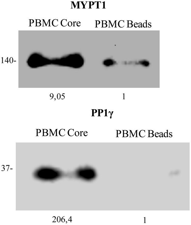
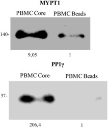
- Figure 4 HCV Core protein interacts with PP1gamma and MYPT1 in PBMC blasts. Postnuclear lysates from Human PBMCs stimulated with PHA and cultured during 5 days with IL-2, were subjected to HCV Core protein pull down. Proteins pulled down by HCV Core coated magnetic beads (PBMC Core band) or by uncoated magnetic beads (PBMC beads band) were analyzed by Western blot with anti-human MYPT1 (A) and anti-human PP1gamma (B). Molecular mass, in kDa, is indicated on the side and band quantitation at the bottom for each Western blot.