Explore
 Validate
 Learn
Learn Western blot
Antibody data
- Antibody Data
- Antigen structure
- References [1]
- Comments [0]
- Validations
- Western blot [1]
- Immunocytochemistry [1]
- Immunohistochemistry [1]
Submit
Validation data
Reference
Comment
Report error
- Product number
- AF7117 - Provider product page

- Provider
- R&D Systems
- Product name
- Human/Mouse MAGI2 Antibody
- Antibody type
- Polyclonal
- Description
- Immunogen affinity purified. Detects human MAGI2 in direct ELISAs and Western blots. In direct ELISAs, less than 1% cross-reactivity with recombinant ARIP-2 is observed.
- Reactivity
- Human, Mouse
- Host
- Goat
- Conjugate
- Unconjugated
- Antigen sequence
Q86UL8- Isotype
- IgG
- Vial size
- 100 ug
- Concentration
- LYOPH
- Storage
- Use a manual defrost freezer and avoid repeated freeze-thaw cycles. 12 months from date of receipt, -20 to -70 °C as supplied. 1 month, 2 to 8 °C under sterile conditions after reconstitution. 6 months, -20 to -70 °C under sterile conditions after reconstitution.
Submitted references Weighted Gene Correlation Network Analysis (WGCNA) Detected Loss of MAGI2 Promotes Chronic Kidney Disease (CKD) by Podocyte Damage.
Zuo Z, Shen JX, Pan Y, Pu J, Li YG, Shao XH, Wang WP
Cellular physiology and biochemistry : international journal of experimental cellular physiology, biochemistry, and pharmacology 2018;51(1):244-261
Cellular physiology and biochemistry : international journal of experimental cellular physiology, biochemistry, and pharmacology 2018;51(1):244-261
No comments: Submit comment
Supportive validation
- Submitted by
- R&D Systems (provider)
- Main image
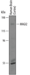
- Experimental details
- Detection of Human MAGI2 by Western Blot. Western blot shows lysates of human brain (cortex) tissue. PVDF membrane was probed with 1 µg/mL of Goat Anti-Human MAGI2 Antigen Affinity-purified Polyclonal Antibody (Catalog # AF7117) followed by HRP-conjugated Anti-Goat IgG Secondary Antibody (Catalog # HAF019). A specific band was detected for MAGI2 at approximately 170 kDa (as indicated). This experiment was conducted under reducing conditions and using Immunoblot Buffer Group 1.
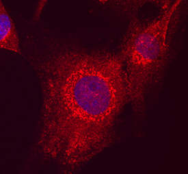
Supportive validation
- Submitted by
- R&D Systems (provider)
- Main image

- Experimental details
- MAGI2 in U-87 MG Human Cell Line. MAGI2 was detected in immersion fixed U-87 MG human glioblastoma/ astrocytoma cell line using Goat Anti-Human MAGI2 Antigen Affinity-purified Polyclonal Antibody (Catalog # AF7117) at 10 µg/mL for 3 hours at room temperature. Cells were stained using the NorthernLights™ 557-conjugated Anti-Goat IgG Secondary Antibody (red; Catalog # NL001) and counterstained with DAPI (blue). Specific staining was localized to cytoplasm. View our protocol for Fluorescent ICC Staining of Cells on Coverslips.
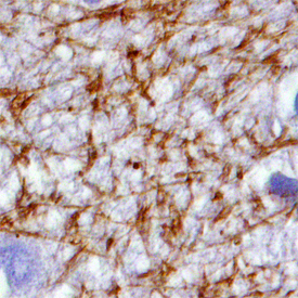
Supportive validation
- Submitted by
- R&D Systems (provider)
- Main image

- Experimental details
- MAGI2 in Human Brain. MAGI2 was detected in immersion fixed paraffin-embedded sections of human brain (hippocampus) using Goat Anti-Human MAGI2 Antigen Affinity-purified Polyclonal Antibody (Catalog # AF7117) at 15 µg/mL overnight at 4 °C. Tissue was stained using the Anti-Goat HRP-DAB Cell & Tissue Staining Kit (brown; Catalog # CTS008) and counterstained with hematoxylin (blue). Specific staining was localized to synaptic boutons and neuronal processes. View our protocol for Chromogenic IHC Staining of Paraffin-embedded Tissue Sections.