Explore
 Validate
 Learn
Learn Western blot
 Immunohistochemistry
 Other assay
Antibody data
- Antibody Data
- Antigen structure
- References [1]
- Comments [0]
- Validations
- Other assay [2]
Submit
Validation data
Reference
Comment
Report error
- Product number
- PA5-36472 - Provider product page

- Provider
- Invitrogen Antibodies
- Product name
- Cerebellin 4 Polyclonal Antibody
- Antibody type
- Polyclonal
- Antigen
- Synthetic peptide
- Description
- This antibody detects endogenous protein at a molecular weight of 27 kDa. Purity is >95% by SDS-PAGE.
- Reactivity
- Human, Mouse, Rat
- Host
- Rabbit
- Isotype
- IgG
- Vial size
- 100 μL
- Concentration
- 1 mg/mL
- Storage
- Store at 4°C short term. For long term storage, store at -20°C, avoiding freeze/thaw cycles.
Submitted references Transcriptional profiling aligned with in situ expression image analysis reveals mosaically expressed molecular markers for GABA neuron sub-groups in the ventral tegmental area.
Paul EJ, Tossell K, Ungless MA
The European journal of neuroscience 2019 Dec;50(11):3732-3749
The European journal of neuroscience 2019 Dec;50(11):3732-3749
No comments: Submit comment
Supportive validation
- Submitted by
- Invitrogen Antibodies (provider)
- Main image
- Experimental details
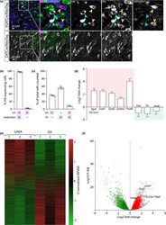
- Figure 1 Isolation and sequencing of RNA from samples enriched for either GABA or dopamine neurons. (a) Representative images showing triple immunolabelling for HA , GABAAR alpha1 and TH in the VTA from a VGATC re:RiboTag mouse (top panels) and double immunofluorescence for HA and TH in the VTA from a DATC re:RiboTag mouse (bottom panels). In each case HA immunoreactivity is selectively localised to the target neuronal population as illustrated by co-localisation with GABAAR alpha1 (in HA +/ TH - cells (yellow arrows) but not in TH + cells (blue arrows)) in the top panels and TH in the bottom panels. (b) Graph showing mean (+ SEM ) percentage of cells ( n = 481) that were either HA +, TH +, or HA + and TH +. (c) Graph showing mean (+ SEM ) percentage of HA + ( TH -) cells ( n = 278) that expressed GABAAR alpha1. (d) Graph showing mean (+ SEM ) GABA neuron-specific transcripts (red box; slc32a1, Gad1, Gad2, Gabra1, Pvalb ) significantly enriched in samples enriched for GABA neurons compared to dopamine neurons ( P-Adj < 0.05). In contrast, dopamine neuron-related transcripts ( green box; Dat, Th, Aadc) were not enriched ( P-Adj > 0.05). (e) Heatmap depicting the 15,015 mRNA transcripts detected in GABA and dopamine neurons (each row represents a different transcript). Expression levels (reads per kilobase of transcript per million mapped reads; RPKM ) were scaled and sorted by z-score. The red-green scale illustrates level of enrichment (red = enriched; green = de-enriched). E
- Submitted by
- Invitrogen Antibodies (provider)
- Main image
- Experimental details
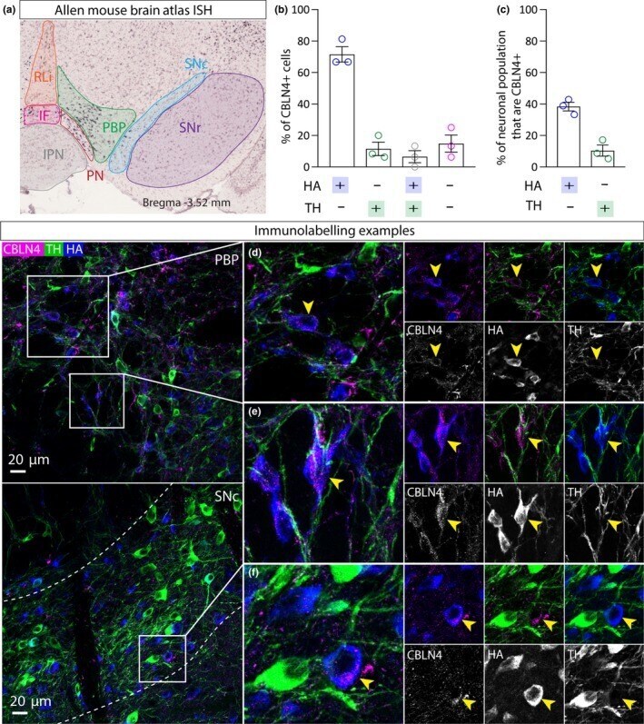
- Figure 4 CBLN 4 expression in GABA neurons in the VTA . a, Left panel shows ISH image from the Allen Mouse Brain Atlas showing mosaic expression of CBLN 4 in the PN and medial PBP . b, Graph showing mean (+ SEM ) percentage of CBLN + cells ( n = 59 cells) that express either HA , or TH , or HA and TH , or neither. c, Graph showing mean (+ SEM ) percentage of HA + cells ( n = 124 cells) that express CBLN and the percentage of TH + cells ( n = 94 cells) that express CBLN . d, e, Representative examples of immunolabelling of CBLN 4 (magenta), HA (blue) and TH (green) in the VTA of a VGATC re:RiboTag mouse, illustrating exemplar cells exhibiting CBLN 4 co-localisation with HA , but not TH (yellow arrows). f, Representative example of immunolabelling in the SN c. [Colour figure can be viewed at http://www.wileyonlinelibrary.com ]