Explore
 Validate
 Learn
LearnPA5-28225
antibody from Invitrogen Antibodies
Targeting: PPP1CB
MP, PP-1B, PP1B, PP1beta, PP1c, PPP1beta
 Western blot
Antibody data
- Antibody Data
- Antigen structure
- References [0]
- Comments [0]
- Validations
- Western blot [5]
- Immunocytochemistry [2]
- Immunohistochemistry [5]
- Chromatin Immunoprecipitation [1]
Submit
Validation data
Reference
Comment
Report error
- Product number
- PA5-28225 - Provider product page

- Provider
- Invitrogen Antibodies
- Product name
- PP1 beta Polyclonal Antibody
- Antibody type
- Polyclonal
- Antigen
- Recombinant protein fragment
- Description
- Recommended positive controls: 293T, A431, HeLa, HepG2, PPP1CB shRNA-transfected 293T, Mouse brain. Predicted reactivity: Mouse (100%), Rat (100%), Zebrafish (99%), Xenopus laevis (100%), Dog (99%), Pig (100%), Rabbit (100%), Chicken (100%), Rhesus Monkey (100%), Bovine (100%). Store product as a concentrated solution. Centrifuge briefly prior to opening the vial.
- Reactivity
- Human, Mouse, Rat
- Host
- Rabbit
- Isotype
- IgG
- Vial size
- 100 µL
- Concentration
- 0.6 mg/mL
- Storage
- Store at 4°C short term. For long term storage, store at -20°C, avoiding freeze/thaw cycles.
No comments: Submit comment
Supportive validation
- Submitted by
- Invitrogen Antibodies (provider)
- Main image

- Experimental details
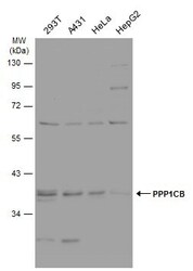
- Western blot analysis of PPP1CB using 30µg of A) 293T (B) A431 (C) H1299 (D) HeLa S3 (E) HepG2 (F) MOLT4 and G) Raji lysate. Samples were loaded onto a 7.5% SDS-PAGE gel and probed with a PPP1CB polyclonal antibody (Product # PA5-28225) at a dilution of 1:1000.

- Submitted by
- Invitrogen Antibodies (provider)
- Main image
- Experimental details
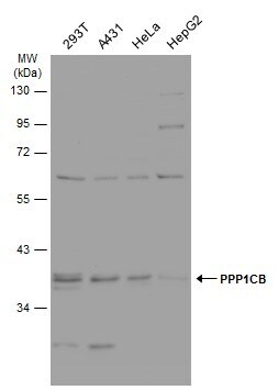
- Western Blot analysis of PP1 beta was performed by separating 30 µg of various whole cell extracts by 10% SDS-PAGE. Proteins were transferred to a membrane and probed with a PP1 beta Polyclonal Antibody (Product # PA5-28225) at a dilution of 1:1000 and a HRP-conjugated anti-rabbit IgG secondary antibody.
- Submitted by
- Invitrogen Antibodies (provider)
- Main image

- Experimental details
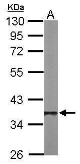
- Western Blot using PP1 beta Polyclonal Antibody (Product # PA5-28225). Sample (50 µg of whole cell lysate). Lane A: Mouse brain. 10% SDS PAGE. PP1 beta Polyclonal Antibody (Product # PA5-28225) diluted at 1:1,000. The HRP-conjugated anti-rabbit IgG antibody was used to detect the primary antibody.
- Submitted by
- Invitrogen Antibodies (provider)
- Main image
- Experimental details
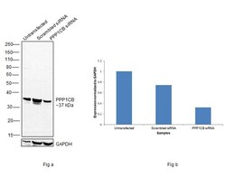
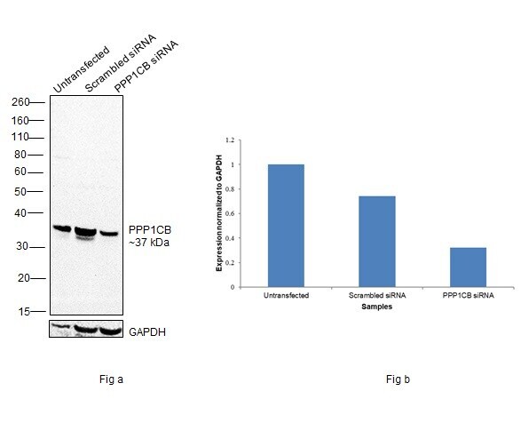
- Knockdown of Serine/threonine-protein phosphatase PP1-beta catalytic subunit was achieved by transfecting HT-29 with Serine/threonine-protein phosphatase PP1-beta catalytic subunit specific siRNAs (Silencer® select Product # S10935, S10934). Western blot analysis (Fig. a) was performed using Membrane enriched extracts from the Serine/threonine-protein phosphatase PP1-beta catalytic subunit knockdown cells (lane 3), non-targeting scrambled siRNA transfected cells (lane 2) and untransfected cells (lane 1). The blot was probed with PP1 beta Polyclonal Antibody (Product # PA5-28225, 1:1000 dilution) and Goat anti-Rabbit IgG (H+L) Superclonal™ Recombinant Secondary Antibody, HRP (Product # A27036,1:4000 dilution). Densitometric analysis of this western blot is shown in histogram (Fig. b). Decrease in signal upon siRNA mediated knock down confirms that antibody is specific to Serine/threonine-protein phosphatase PP1-beta catalytic subunit.
- Submitted by
- Invitrogen Antibodies (provider)
- Main image
- Experimental details
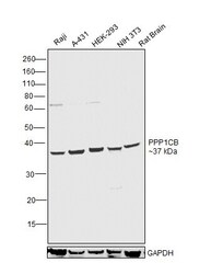
- Western blot was performed using Anti-PP1 beta Polyclonal Antibody (Product # PA5-28225) and a 37 kDa band corresponding to Serine/threonine-protein phosphatase PP1-beta catalytic subunit was observed across cell lines and tissue tested. Whole cell extracts (30 µg lysate) of Raji (Lane 1), A-431 (Lane 2), HEK-293 (Lane 3), NIH 3T3 (Lane 4) and tissue extracts of Rat Brain (Lane 5) were electrophoresed using NuPAGE™ 10% Bis-Tris Protein Gel (Product # NP0302BOX). Resolved proteins were then transferred onto a Nitrocellulose membrane (Product # IB23001) by iBlot® 2 Dry Blotting System (Product # IB21001). The blot was probed with the primary antibody (1:1000 dilution) and detected by chemiluminescence with Goat anti-Rabbit IgG (H+L) Superclonal™ Recombinant Secondary Antibody, HRP (Product # A27036, 1:4000 dilution) using the iBright FL 1000 (Product # A32752). Chemiluminescent detection was performed using Novex® ECL Reagent Kit (Product # WP20005).

Supportive validation
- Submitted by
- Invitrogen Antibodies (provider)
- Main image
- Experimental details
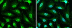
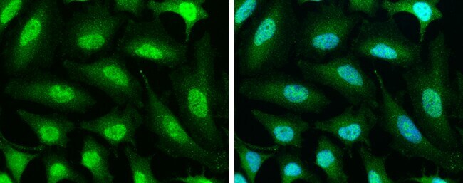
- Immunocytochemistry-Immunofluorescence analysis of PP1 beta was performed in HeLa cells fixed in 4% paraformaldehyde at RT for 15 min. Green: PP1 beta Polyclonal Antibody (Product # PA5 28225) diluted at 1:500. Blue: Hoechst 33342 staining.
- Submitted by
- Invitrogen Antibodies (provider)
- Main image
- Experimental details
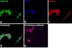
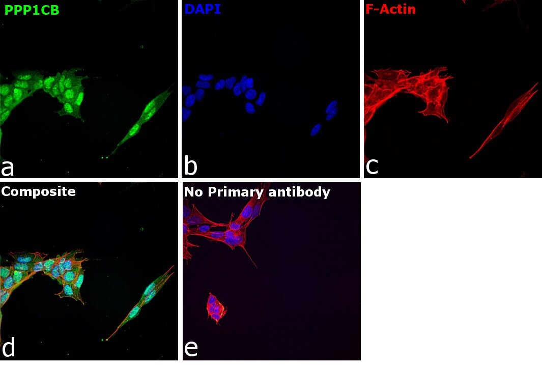
- Immunofluorescence analysis of PPP1CB was performed using 70% confluent log phase SH-SY5Y cells. The cells were fixed with 4% paraformaldehyde for 10 minutes, permeabilized with 0.1% Triton™ X-100 for 15 minutes, and blocked with 2% BSA for 1 hour at room temperature. The cells were labeled with PP1 beta Polyclonal Antibody (Product # PA5-28225) at 1:100 dilution in 0.1% BSA, incubated at 4 degree celsius overnight and then with Donkey anti-Rabbit IgG (H+L) Highly Cross-Adsorbed Secondary Antibody, Alexa Fluor Plus 488 (Product # A32790) at a dilution of 1:2000 dilution for 45 minutes at room temperature (Panel a: green). Nuclei (Panel b: blue) were stained with Hoechst 33342 (Product # H1399). F-actin (Panel c: red) was stained with Rhodamine Phalloidin (Product # R415, 1:300 dilution). Panel d represents the merged image showing cytoplasmic and nucleus localization. Panel e represents control cells with no primary antibody to assess background. The images were captured at 40X magnification in CellInsight CX7 LZR High-Content Screening (HCS) Platform (Product # CX7C1115LZR).
Supportive validation
- Submitted by
- Invitrogen Antibodies (provider)
- Main image

- Experimental details
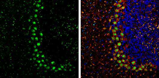
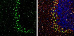
- Immunohistochemistry (Frozen) analysis of PP1-beta was performed in frozen-sectioned adult mouse cerebellum tissue using PP1 beta Polyclonal Antibody (Product # PA5-28225) at a dilution of 1:250 (Green). Red: NF-H, stained by NF-H antibody diluted at 1:500. Blue: Fluoroshield with DAPI. Antigen Retrieval: Citrate buffer, pH 6.0, 10 min.
- Submitted by
- Invitrogen Antibodies (provider)
- Main image
- Experimental details
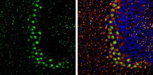
- Immunohistochemistry (Frozen) analysis of PP1-beta was performed in frozen-sectioned adult mouse cerebellum tissue using PP1 beta Polyclonal Antibody (Product # PA5-28225) at a dilution of 1:250 (Green). Red: NF-H, stained by NF-H antibody diluted at 1:500. Blue: Fluoroshield with DAPI. Antigen Retrieval: Citrate buffer, pH 6.0, 10 min.
- Submitted by
- Invitrogen Antibodies (provider)
- Main image

- Experimental details
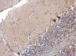
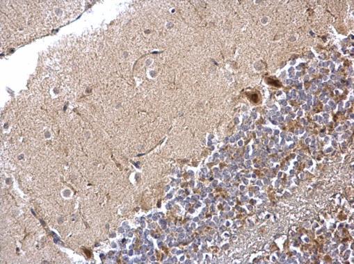
- PP1 beta Polyclonal Antibody detects PPP1CB protein at cytoplasm and nucleus by immunohistochemical analysis. Sample: Paraffin-embedded mouse brain. PPP1CB stained by PP1 beta Polyclonal Antibody (Product # PA5-28225) diluted at 1:3,000. Antigen Retrieval: Citrate buffer, pH 6.0, 15 min.

- Submitted by
- Invitrogen Antibodies (provider)
- Main image
- Experimental details
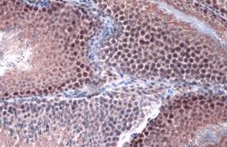
- PP1 beta Polyclonal Antibody detects PPP1CB protein at cytoplasm and nucleus by immunohistochemical analysis. Sample: Paraffin-embedded rat testis. PPP1CB stained by PP1 beta Polyclonal Antibody (Product # PA5-28225) diluted at 1:3,000. Antigen Retrieval: Citrate buffer, pH 6.0, 15 min.

- Submitted by
- Invitrogen Antibodies (provider)
- Main image
- Experimental details
- PP1 beta Polyclonal Antibody detects PPP1CB protein at nucleus and cytosol on mouse lung by immunohistochemical analysis. Sample: Paraffin-embedded mouse lung. PP1 beta Polyclonal Antibody (Product # PA5-28225) dilution: 1:500. Antigen Retrieval: EDTA based buffer, pH 8.0, 15 min.
Supportive validation
- Submitted by
- Invitrogen Antibodies (provider)
- Main image

- Experimental details
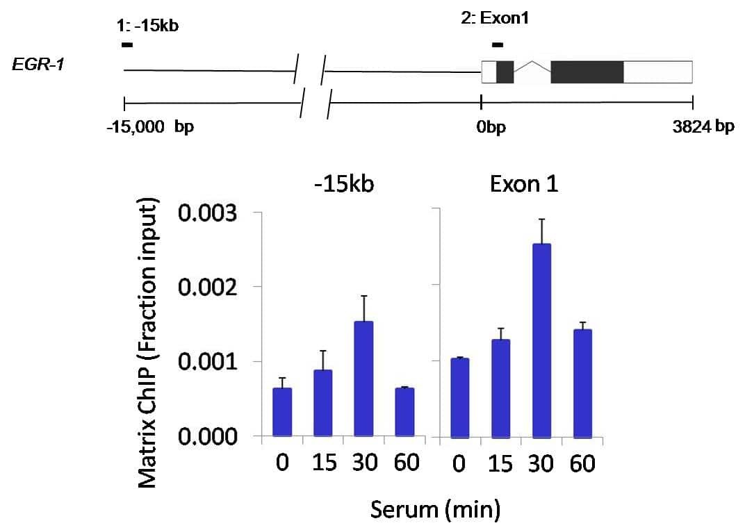
- Chromatin immunoprecipitation analysis of PPP1CB was performed using cross-linked chromatin from 1x10^6 HCT116 colon carcinoma cells treated with serum for 0, 15, 30, and 60 minutes. Immunoprecipitation was performed using a multiplex microplate Matrix ChIP assay (see reference for Matrix ChIP protocol: http://www.ncbi.nlm.nih.gov/pubmed/22098709) with 1.0 µL/100 µL well volume of a PPP1CB polyclonal antibody (Product # PA5-28225). Chromatin aliquots from ~1 x 10⁵ cells were used per ChIP pull-down. Quantitative PCR data were done in quadruplicate using 1 µL of eluted DNA in 2 µL SYBR real-time PCR reactions containing primers to amplify -15kb upstream of the Egr1 gene or exon-1 of Egr1. PCR calibration curves were generated for each primer pair from a dilution series of sheared total genomic DNA. Quantitation of immunoprecipitated chromatin is presented as signal relative to the total amount of input chromatin. Results represent the mean +/- SEM for three experiments. A schematic representation of the Egr-1 locus is shown above the data where boxes represent exons (black boxes = translated regions, white boxes = untranslated regions), the zigzag line represents an intron, and the straight line represents upstream sequence. Regions amplified by Egr-1 primers are represented by black bars. Data courtesy of the Innovators Program.