Explore
 Validate
 Learn
Learn Western blot
 Immunoprecipitation
Antibody data
- Antibody Data
- Antigen structure
- References [6]
- Comments [0]
- Validations
- Western blot [1]
- Immunocytochemistry [4]
- Immunohistochemistry [1]
- Other assay [2]
Submit
Validation data
Reference
Comment
Report error
- Product number
- PA3-910 - Provider product page

- Provider
- Invitrogen Antibodies
- Product name
- GRASP65 Polyclonal Antibody
- Antibody type
- Polyclonal
- Antigen
- Other
- Description
- PA3-910 detects Grasp65 from human, mouse, and rat samples. PA3-910 has been successfully used in Western blot, IHC (paraffin), immunofluorescence, immunocytochemistry and immunoprecipitation procedures. The PA3-910 immunogen is a GST fustion protein containing the C-terminal residues (202-447) of rat Grasp65.
- Reactivity
- Human, Mouse, Rat
- Host
- Rabbit
- Isotype
- IgG
- Vial size
- 100 µL
- Concentration
- Conc. Not Determined
- Storage
- -20° C, Avoid Freeze/Thaw Cycles
Submitted references Lack of N-glycosylation increases amyloidogenic processing of the amyloid precursor protein.
Dynamin Inhibitors Prevent the Establishment of the Cytomegalovirus Assembly Compartment in the Early Phase of Infection.
Towards Age-Related Anti-Inflammatory Therapy: Klotho Suppresses Activation of ER and Golgi Stress Response in Senescent Monocytes.
Nanofibrous Tubular Three-Dimensional Platform for Single Dental Pulp Stem Cell Polarization.
Serotonin 5-HT(2C) Receptor Cys23Ser Single Nucleotide Polymorphism Associates with Receptor Function and Localization In Vitro.
Autophagy-lysosome pathway alterations and alpha-synuclein up-regulation in the subtype of neuronal ceroid lipofuscinosis, CLN5 disease.
Lin T, van Husen LS, Yu Y, Tjernberg LO, Schedin-Weiss S
Glycobiology 2022 May 23;32(6):506-517
Glycobiology 2022 May 23;32(6):506-517
Dynamin Inhibitors Prevent the Establishment of the Cytomegalovirus Assembly Compartment in the Early Phase of Infection.
Štimac I, Jug Vučko N, Blagojević Zagorac G, Marcelić M, Mahmutefendić Lučin H, Lučin P
Life (Basel, Switzerland) 2021 Aug 25;11(9)
Life (Basel, Switzerland) 2021 Aug 25;11(9)
Towards Age-Related Anti-Inflammatory Therapy: Klotho Suppresses Activation of ER and Golgi Stress Response in Senescent Monocytes.
Mytych J, Sołek P, Będzińska A, Rusinek K, Warzybok A, Tabęcka-Łonczyńska A, Koziorowski M
Cells 2020 Jan 21;9(2)
Cells 2020 Jan 21;9(2)
Nanofibrous Tubular Three-Dimensional Platform for Single Dental Pulp Stem Cell Polarization.
Chang B, Ma C, Liu X
ACS applied materials & interfaces 2020 Dec 9;12(49):54481-54488
ACS applied materials & interfaces 2020 Dec 9;12(49):54481-54488
Serotonin 5-HT(2C) Receptor Cys23Ser Single Nucleotide Polymorphism Associates with Receptor Function and Localization In Vitro.
Land MA, Chapman HL, Davis-Reyes BD, Felsing DE, Allen JA, Moeller FG, Elferink LA, Cunningham KA, Anastasio NC
Scientific reports 2019 Nov 13;9(1):16737
Scientific reports 2019 Nov 13;9(1):16737
Autophagy-lysosome pathway alterations and alpha-synuclein up-regulation in the subtype of neuronal ceroid lipofuscinosis, CLN5 disease.
Adams J, Feuerborn M, Molina JA, Wilden AR, Adhikari B, Budden T, Lee SY
Scientific reports 2019 Jan 17;9(1):151
Scientific reports 2019 Jan 17;9(1):151
No comments: Submit comment
Supportive validation
- Submitted by
- Invitrogen Antibodies (provider)
- Main image
- Experimental details
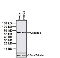
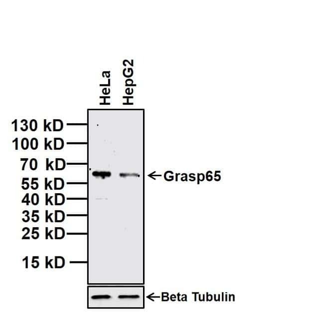
- Western blot analysis of Grasp65 was performed by loading 20 µg of indicated whole cell lysates and 7 µL of PageRuler Prestained Protein Ladder (Product # 26616) per well onto a 4-20% Tris-Glycine polyacrylamide gel (Product # WT4202BX10). Proteins were transferred to a nitrocellulose membrane using the G2 Blotter (Product # 62288), and blocked with 5% Milk in TBST for 1 hour at room temperature. Grasp65 was detected at ~65 kDa using a rabbit polyclonal antibody (Product # PA3-910, upper panel) at a dilution of 1:5000 in blocking buffer overnight at 4°C on a rocking platform ), followed by a Goat anti-Rabbit IgG (H+L) Secondary Antibody, HRP conjugate (Product # 31460) at a dilution of 1:20000 for at least 30 minutes at room temperature. Beta Tubulin was detected using Beta Tubulin mouse monoclonal antibody (Product # MA5-16308, lower panel), followed by a Goat anti-Mouse IgG (H+L) Secondary Antibody, HRP conjugate (Product # 31430) at a dilution of 1:20000 for at least 30 minutes at room temperature. Chemiluminescent detection was performed using SuperSignal West Dura (Product # 34076).
Supportive validation
- Submitted by
- Invitrogen Antibodies (provider)
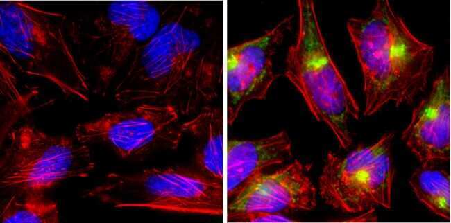
- Main image
- Experimental details
- Immunofluorescent analysis of GRASP65 (green) showing staining in the Golgi apparatus of HeLa cells. Formalin-fixed cells were permeabilized with 0.1% Triton X-100 in TBS for 5-10 minutes and blocked with 3% BSA-PBS for 30 minutes at room temperature. Cells were probed with a GRASP65 polyclonal antibody (Product # PA3-910) in 3% BSA-PBS at a dilution of 1:200 and incubated overnight at 4 ºC in a humidified chamber. Cells were washed with PBST and incubated with a DyLight-conjugated secondary antibody in PBS at room temperature in the dark. F-actin (red) was stained with a fluorescent red phalloidin and nuclei (blue) were stained with Hoechst or DAPI. Images were taken at a magnification of 100x.
- Submitted by
- Invitrogen Antibodies (provider)
- Main image
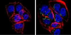
- Experimental details
- Immunofluorescent analysis of GRASP65 (green) showing staining in the Golgi apparatus of HepG2 cells. Formalin-fixed cells were permeabilized with 0.1% Triton X-100 in TBS for 5-10 minutes and blocked with 3% BSA-PBS for 30 minutes at room temperature. Cells were probed with a GRASP65 polyclonal antibody (Product # PA3-910) in 3% BSA-PBS at a dilution of 1:200 and incubated overnight at 4 ºC in a humidified chamber. Cells were washed with PBST and incubated with a DyLight-conjugated secondary antibody in PBS at room temperature in the dark. F-actin (red) was stained with a fluorescent red phalloidin and nuclei (blue) were stained with Hoechst or DAPI. Images were taken at a magnification of 100x.

- Submitted by
- Invitrogen Antibodies (provider)
- Main image
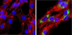
- Experimental details
- Immunofluorescent analysis of GRASP65 (green) showing staining in the Golgi apparatus of PC12 cells. Formalin-fixed cells were permeabilized with 0.1% Triton X-100 in TBS for 5-10 minutes and blocked with 3% BSA-PBS for 30 minutes at room temperature. Cells were probed with a GRASP65 polyclonal antibody (Product # PA3-910) in 3% BSA-PBS at a dilution of 1:200 and incubated overnight at 4 ºC in a humidified chamber. Cells were washed with PBST and incubated with a DyLight-conjugated secondary antibody in PBS at room temperature in the dark. F-actin (red) was stained with a fluorescent red phalloidin and nuclei (blue) were stained with Hoechst or DAPI. Images were taken at a magnification of 100x.
- Submitted by
- Invitrogen Antibodies (provider)
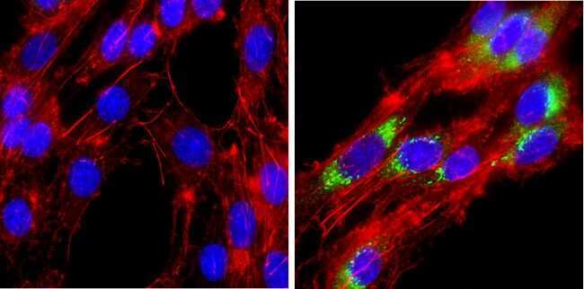
- Main image
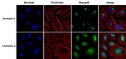
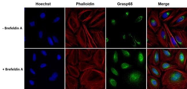
- Experimental details
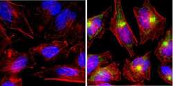
- Immunofluorescent analysis of Grasp65 (green) in HeLa cells either left untreated or treated 5 µg/mL Brefeldin A for 4 hours. The cells were fixed with 4% paraformaldehyde for 15 minutes at room temperature, permeabilized with 0.1% Triton X-100 for 15 minutes, and blocked with 3% BSA for 30 minutes at room temperature. Cells were stained with a Grasp65 rabbit polyclonal antibody (Product # PA3-910) at a dilution of 1:200 in blocking buffer for 1 hour at room temperature, and then incubated with a Goat anti-Rabbit IgG (H+L) Secondary Antibody, Alexa Fluor Plus 488 conjugate (Product # A32731) at a dilution of 1:500 for at least 30 minutes at a room temperature in the dark (green). F-actin (red) was stained with Dylight 554 Phalloidin. Nuclei (blue) were stained with Hoechst 33342 (Product # 62249). Images were taken on a Thermo Scientific ToxInsight Instrument at 20X magnification.
Supportive validation
- Submitted by
- Invitrogen Antibodies (provider)
- Main image
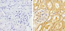
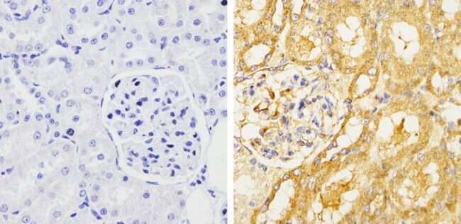
- Experimental details
- Immunohistochemistry analysis of GRASP65 showing staining in the cytoplasm of paraffin-treated rat kidney tissue (right) compared with a negative control without primary antibody (left). To expose target proteins, antigen retrieval was performed using 10mM sodium citrate (pH 6.0), microwaved for 8-15 min. Following antigen retrieval, tissues were blocked in 3% H2O2-methanol for 15 min at room temperature, washed with ddH2O and PBS, and then probed with a GRASP65 polyclonal antibody (Product # PA3-910) diluted by 3% BSA-PBS at a dilution of 1:200 overnight at 4°C in a humidified chamber. Tissues were washed extensively in PBST and detection was performed using an HRP-conjugated secondary antibody followed by colorimetric detection using a DAB kit. Tissues were counterstained with hematoxylin and dehydrated with ethanol and xylene to prep for mounting.
Supportive validation
- Submitted by
- Invitrogen Antibodies (provider)
- Main image
- Experimental details
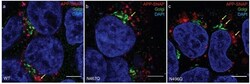
- Localization of APP-SNAP variants in proximity to the Golgi apparatus. Confocal images of APP-SNAP variants in proximity to the Golgi apparatus, imaged 48 h after transfection of HEK293T cells. Representative images for WT (a), N467QA (b), and N496Q (c) are shown. Anti-GRASP65 antibody was used to stain the Golgi apparatus, while the SNAP-tag of APP was labelled with TMR-star (red) and DAPI was used to stain nuclei (blue). Yellow arrows point at vesicles that are sufficiently resolved to show the location of APP in the vesicular membrane. Scale bars: 5 mum.

- Submitted by
- Invitrogen Antibodies (provider)
- Main image
- Experimental details
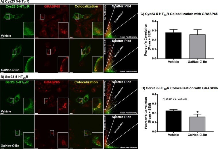
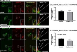
- Figure 4 GalNac- O -Bn treatment decreases colocalization of the Ser23 allele with GRASP65 with no effect on the Cys23 allele. Colocalization of transiently transfected CHOp38 cells expressing (A) Cys23 or (B) Ser23 5-HT 2C R in green with the Golgi reassembly-stacking protein of 65 kDa (GRASP65) in red treated with vehicle or 2 mM Benzyl 2-acetamido-2-deoxy-alpha-D-galactopyranoside (GalNAc- O -Bn) and represented as a single mid-cell slice. The scatter plot represents the type and intensity of pixels for the evaluated cell. Scale bar = 10 um. Pearson's Correlation between the 5-HT 2C R and the GRASP65 for (C) Cys23 and (D) Ser23 CHOp38 cells. Each experiment was performed on four to eight biological replicates with 20-30 fields of view per experiment and 40-70 cells analyzed for each condition per receptor variant. *p < 0.05 vs. Vehicle.