Explore
 Validate
 Learn
Learn Immunohistochemistry
Antibody data
- Antibody Data
- Antigen structure
- References [5]
- Comments [0]
- Validations
- Immunohistochemistry [1]
Submit
Validation data
Reference
Comment
Report error
- Product number
- HPA014067 - Provider product page

- Provider
- Atlas Antibodies
- Proper citation
- Atlas Antibodies Cat#HPA014067, RRID:AB_1848178
- Product name
- Anti-ENTPD1
- Antibody type
- Polyclonal
- Description
- Polyclonal Antibody against Human ENTPD1, Gene description: ectonucleoside triphosphate diphosphohydrolase 1, Alternative Gene Names: ATPDase, CD39, NTPDase-1, SPG64, Validated applications: IHC, Uniprot ID: P49961, Storage: Store at +4°C for short term storage. Long time storage is recommended at -20°C.
- Reactivity
- Human
- Host
- Rabbit
- Conjugate
- Unconjugated
- Isotype
- IgG
- Vial size
- 100 µl
- Concentration
- 0.05 mg/ml
- Storage
- Store at +4°C for short term storage. Long time storage is recommended at -20°C.
- Handling
- The antibody solution should be gently mixed before use.
Submitted references The expression of the adenosine pathway markers CD39 and CD73 in salivary gland carcinomas harbors the potential for novel immune checkpoint inhibition
Limited TCR repertoire and ENTPD1 dysregulation mark late-stage COVID-19
CD39 is a promising therapeutic antibody target for the treatment of soft tissue sarcoma.
Impaired natriuretic response to high-NaCl diet plus aldosterone infusion in mice overexpressing human CD39, an ectonucleotidase (NTPDase1)
Simultaneous Overexpression of Functional Human HO-1, E5NT and ENTPD1 Protects Murine Fibroblasts against TNF-α-Induced Injury In Vitro
Bauer A, Gebauer N, Knief J, Tharun L, Arnold N, Riecke A, Steinestel K, Witte H
Journal of Cancer Research and Clinical Oncology 2022;149(7):3193-3208
Journal of Cancer Research and Clinical Oncology 2022;149(7):3193-3208
Limited TCR repertoire and ENTPD1 dysregulation mark late-stage COVID-19
Wang N, Vuerich M, Kalbasi A, Graham J, Csizmadia E, Manickas-Hill Z, Woolley A, David C, Miller E, Gorman K, Hecht J, Shaefi S, Robson S, Longhi M
iScience 2021;24(10):103205
iScience 2021;24(10):103205
CD39 is a promising therapeutic antibody target for the treatment of soft tissue sarcoma.
Hayes GM, Cairns B, Levashova Z, Chinn L, Perez M, Theunissen JW, Liao-Chan S, Bermudez A, Flory MR, Schweighofer KJ, H van der Horst E
American journal of translational research 2015;7(6):1181-8
American journal of translational research 2015;7(6):1181-8
Impaired natriuretic response to high-NaCl diet plus aldosterone infusion in mice overexpressing human CD39, an ectonucleotidase (NTPDase1)
Zhang Y, Robson S, Morris K, Heiney K, Dwyer K, Kishore B, Ecelbarger C
American Journal of Physiology-Renal Physiology 2015;308(12):F1398-F1408
American Journal of Physiology-Renal Physiology 2015;308(12):F1398-F1408
Simultaneous Overexpression of Functional Human HO-1, E5NT and ENTPD1 Protects Murine Fibroblasts against TNF-α-Induced Injury In Vitro
Slominski A, Cinti A, De Giorgi M, Chisci E, Arena C, Galimberti G, Farina L, Bugarin C, Rivolta I, Gaipa G, Smolenski R, Cerrito M, Lavitrano M, Giovannoni R
PLOS ONE 2015;10(10):e0141933
PLOS ONE 2015;10(10):e0141933
No comments: Submit comment
Supportive validation
- Submitted by
- Atlas Antibodies (provider)
- Enhanced method
- Orthogonal validation
- Main image
- Experimental details
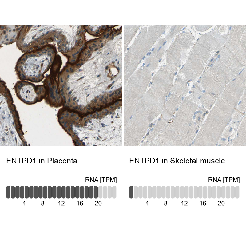
- Immunohistochemistry analysis in human placenta and skeletal muscle tissues using HPA014067 antibody. Corresponding ENTPD1 RNA-seq data are presented for the same tissues.
- Sample type
- Human
- Protocol
- Protocol