Explore
 Validate
 Learn
Learn Western blot
 ELISA
ELISAAntibody data
- Antibody Data
- Antigen structure
- References [11]
- Comments [0]
- Validations
- Western blot [1]
- Immunocytochemistry [1]
- Immunohistochemistry [2]
Submit
Validation data
Reference
Comment
Report error
- Product number
- 60063-1-Ig - Provider product page

- Provider
- Proteintech Group
- Proper citation
- Proteintech Cat#60063-1-Ig, RRID:AB_2197544
- Product name
- Stanniocalcin 2 antibody
- Antibody type
- Monoclonal
- Description
- KD/KO validated Stanniocalcin 2 antibody (Cat. #60063-1-Ig) is a mouse monoclonal antibody that shows reactivity with human, mouse, pig and has been validated for the following applications: IF, IHC, WB,ELISA.
- Reactivity
- Human, Mouse, Porcine
- Host
- Mouse
- Conjugate
- Unconjugated
- Isotype
- IgG
- Antibody clone number
- 4H1E7
- Vial size
- 20ul, 150ul
Submitted references Smoking-Induced STC2(+) Tumor Cells Drive Tumor-Vascular Crosstalk in Laryngeal Squamous Cell Carcinoma via Spatial and Single-Cell Transcriptomics.
A novel identified epithelial ligand-receptor-associated gene signature highlights POPDC3 as a potential therapy target for non-small cell lung cancer.
STC2 Serves as a Critical Hypoxic Effector in Keloid Pathogenesis by Orchestrating Fibroblasts Activation and ECM Remodelling.
FUT3 mediated GRP78 fucosylation promotes colorectal cancer survival and proliferation under glucose restriction via PERK/ATF4/STC2 axis.
STC2 knockdown inhibits cell proliferation and glycolysis in hepatocellular carcinoma through promoting autophagy by PI3K/Akt/mTOR pathway.
Osteosarcoma transcriptome data exploration reveals STC2 as a novel risk indicator in disease progression.
The Oncogenic and Diagnostic Potential of Stanniocalcin 2 in Hepatocellular Carcinoma.
The novel prognostic risk factor STC2 can regulate the occurrence and progression of osteosarcoma via the glycolytic pathway.
STC2 Is a Potential Prognostic Biomarker for Pancreatic Cancer and Promotes Migration and Invasion by Inducing Epithelial-Mesenchymal Transition.
Profiling the Gene Expression and DNA Methylation in the Mouse Brain after Ischemic Preconditioning.
STC2 promotes head and neck squamous cell carcinoma metastasis through modulating the PI3K/AKT/Snail signaling.
Shen Y, Gao L, Huang Q, Zhou L, Xu C, Wu C
Advanced science (Weinheim, Baden-Wurttemberg, Germany) 2026 Jan;13(5):e11932
Advanced science (Weinheim, Baden-Wurttemberg, Germany) 2026 Jan;13(5):e11932
A novel identified epithelial ligand-receptor-associated gene signature highlights POPDC3 as a potential therapy target for non-small cell lung cancer.
Zhu XR, Zhu JQ, Gu QH, Liu N, Lu JJ, Li XH, Liu YY, Zheng X, Chen MB, Ji Y
Cell death & disease 2025 Feb 19;16(1):114
Cell death & disease 2025 Feb 19;16(1):114
STC2 Serves as a Critical Hypoxic Effector in Keloid Pathogenesis by Orchestrating Fibroblasts Activation and ECM Remodelling.
Qian L, Deng S, Tian T, He Y, Li Y, Wang X, Tian T, Jiang M, Pan Y, Li D
Experimental dermatology 2025 Dec;34(12):e70193
Experimental dermatology 2025 Dec;34(12):e70193
FUT3 mediated GRP78 fucosylation promotes colorectal cancer survival and proliferation under glucose restriction via PERK/ATF4/STC2 axis.
Qin K, Zhang H, Shi Y, Wu R, Wu C, Wu Y, He C, Han Z, Luo Y, Fang Y, Yan X, Wang X, Li M, Li A, Liu S, Li Q
NPJ precision oncology 2025 Dec 14;10(1):19
NPJ precision oncology 2025 Dec 14;10(1):19
STC2 knockdown inhibits cell proliferation and glycolysis in hepatocellular carcinoma through promoting autophagy by PI3K/Akt/mTOR pathway.
Li D, Xiong Y, Li M, Long L, Zhang Y, Yan H, Xiang H
Archives of biochemistry and biophysics 2024 Nov;761:110149
Archives of biochemistry and biophysics 2024 Nov;761:110149
Osteosarcoma transcriptome data exploration reveals STC2 as a novel risk indicator in disease progression.
Wang Z, Zeng Z, Gao F, Gui Z, Du J, Shen N, Shang Y, Yang Z, Shang L, Wei R, Ma W, Wang C
BMC medical genomics 2023 Feb 20;16(1):30
BMC medical genomics 2023 Feb 20;16(1):30
The Oncogenic and Diagnostic Potential of Stanniocalcin 2 in Hepatocellular Carcinoma.
Wu Z, Cheng H, Liu J, Zhang S, Zhang M, Liu F, Li Y, Huang Q, Jiang Y, Chen S, Lv L, Li D, Zeng JZ
Journal of hepatocellular carcinoma 2022;9:141-155
Journal of hepatocellular carcinoma 2022;9:141-155
The novel prognostic risk factor STC2 can regulate the occurrence and progression of osteosarcoma via the glycolytic pathway.
Yu B, Zhang F, Liu L, Liang Y, Tang X, Peng Y, Cai F, Zeng D, Yuan X, Li J, Guo Y, Lv B, Wang M, Liao Q, Lv XB
Biochemical and biophysical research communications 2021 May 21;554:25-32
Biochemical and biophysical research communications 2021 May 21;554:25-32
STC2 Is a Potential Prognostic Biomarker for Pancreatic Cancer and Promotes Migration and Invasion by Inducing Epithelial-Mesenchymal Transition.
Lin C, Sun L, Huang S, Weng X, Wu Z
BioMed research international 2019;2019:8042489
BioMed research international 2019;2019:8042489
Profiling the Gene Expression and DNA Methylation in the Mouse Brain after Ischemic Preconditioning.
Cai M, Zhu Y, Li Z, Josephs-Spaulding J, Zhou Y, Hu Y, Chen H, Liu Y, He W, Zhang J
Neuroscience 2019 May 15;406:249-261
Neuroscience 2019 May 15;406:249-261
STC2 promotes head and neck squamous cell carcinoma metastasis through modulating the PI3K/AKT/Snail signaling.
Yang S, Ji Q, Chang B, Wang Y, Zhu Y, Li D, Huang C, Wang Y, Sun G, Zhang L, Guan Q, Xiang J, Wei W, Lu Z, Liao T, Meng J, Wang Z, Ma B, Zhou L, Wang Y, Yang G
Oncotarget 2017 Jan 24;8(4):5976-5991
Oncotarget 2017 Jan 24;8(4):5976-5991
No comments: Submit comment
Supportive validation
- Submitted by
- Proteintech Group (provider)
- Main image

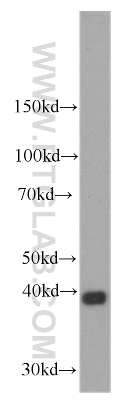
- Experimental details
- HEK-293 cells were subjected to SDS PAGE followed by western blot with 60063-1-Ig(STC2 antibody) at dilution of 1:1000
- Sample type
- cell line
Supportive validation
- Submitted by
- Proteintech Group (provider)
- Main image
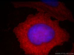
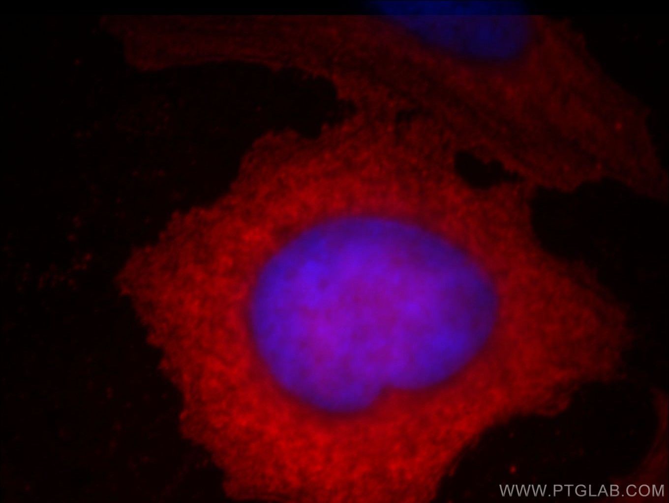
- Experimental details
- Immunofluorescent analysis of Hela cells, using STC2 antibody 60063-1-lg at 1:25 dilution and Rhodamine-labeled goat anti-mouse IgG (red). Blue pseudocolor = DAPI (fluorescent DNA dye).
- Sample type
- cell line
Supportive validation
- Submitted by
- Proteintech Group (provider)
- Main image
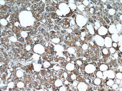
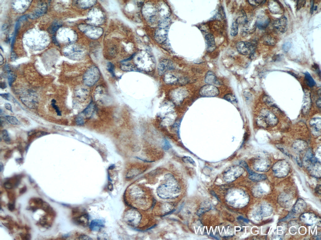
- Experimental details
- Immunohistochemical of paraffin-embedded human breast cancer using 60063-1-Ig(STC2 antibody) at dilution of 1:50 (under 10x lens)
- Sample type
- tissue
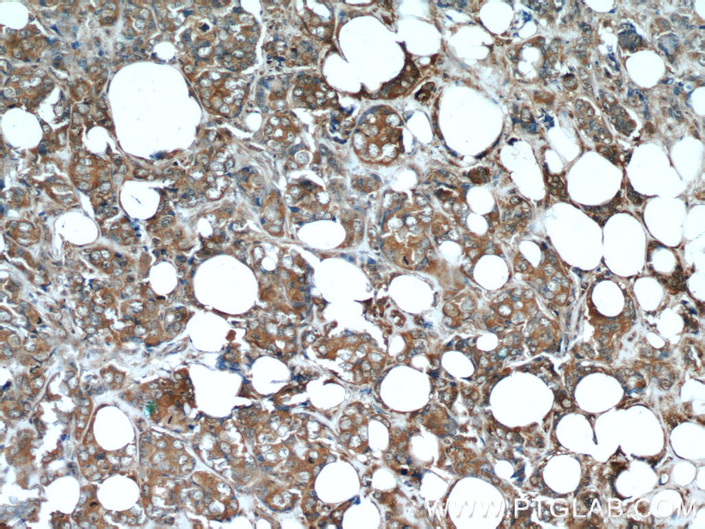
- Submitted by
- Proteintech Group (provider)
- Main image

- Experimental details
- Immunohistochemical of paraffin-embedded human breast cancer using 60063-1-Ig(STC2 antibody) at dilution of 1:50 (under 40x lens)
- Sample type
- tissue