Explore
 Validate
 Learn
Learn Western blot
 Immunocytochemistry
 Immunoprecipitation
Antibody data
- Antibody Data
- Antigen structure
- References [1]
- Comments [0]
- Validations
- Immunocytochemistry [7]
- Other assay [1]
Submit
Validation data
Reference
Comment
Report error
- Product number
- MA1-846 - Provider product page

- Provider
- Invitrogen Antibodies
- Product name
- PRC1 Monoclonal Antibody (16F2)
- Antibody type
- Monoclonal
- Antigen
- Recombinant full-length protein
- Description
- MA1-846 detects PRC1 from human, mouse, and rat samples. MA1-846 has been successfully used in Western blot, immunoprecipitation, and immunofluorescence procedures. The MA1-846 immunogen is recombinant PRC1 protein.
- Reactivity
- Human, Mouse, Rat
- Host
- Mouse
- Isotype
- IgG
- Antibody clone number
- 16F2
- Vial size
- 100 μg
- Concentration
- 1 mg/mL
- Storage
- -20°C, Avoid Freeze/Thaw Cycles
Submitted references Novel role for the midbody in primary ciliogenesis by polarized epithelial cells.
Bernabé-Rubio M, Andrés G, Casares-Arias J, Fernández-Barrera J, Rangel L, Reglero-Real N, Gershlick DC, Fernández JJ, Millán J, Correas I, Miguez DG, Alonso MA
The Journal of cell biology 2016 Aug 1;214(3):259-73
The Journal of cell biology 2016 Aug 1;214(3):259-73
No comments: Submit comment
Supportive validation
- Submitted by
- Invitrogen Antibodies (provider)
- Main image
- Experimental details
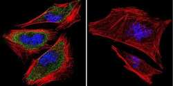
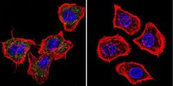
- Immunofluorescent analysis of PRC1 using PRC1 Monoclonal Antibody (16F2) (Product # MA1-846) shows staining in A2058 Cells. PRC1 (green), F-Actin staining with Phalloidin (red) and nuclei with DAPI (blue) is shown. Cells were grown on chamber slides and fixed with formaldehyde prior to staining. Cells were probed without (control) or with an antibody recognizing PRC1 (Product # MA1-846) at a dilution of 1:100 over night at 4 °C, washed with PBS and incubated with a DyLight-488 conjugated secondary antibody (Product # 35552 for GAR, Product # 35503 for GAM). Images were taken at 60X magnification.
- Submitted by
- Invitrogen Antibodies (provider)
- Main image

- Experimental details
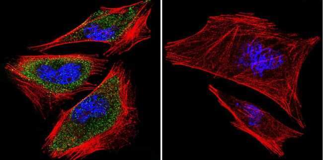
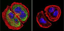
- Immunofluorescent analysis of PRC1 using PRC1 Monoclonal Antibody (16F2) (Product # MA1-846) shows staining in Hela Cells. PRC1 (green), F-Actin staining with Phalloidin (red) and nuclei with DAPI (blue) is shown. Cells were grown on chamber slides and fixed with formaldehyde prior to staining. Cells were probed without (control) or with an antibody recognizing PRC1 (Product # MA1-846) at a dilution of 1:100 over night at 4 °C, washed with PBS and incubated with a DyLight-488 conjugated secondary antibody (Product # 35552 for GAR, Product # 35503 for GAM). Images were taken at 60X magnification.
- Submitted by
- Invitrogen Antibodies (provider)
- Main image
- Experimental details
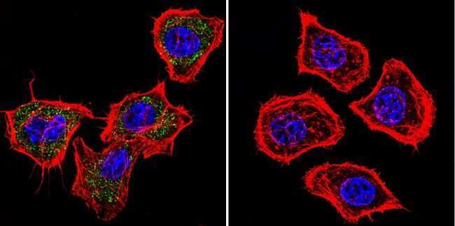
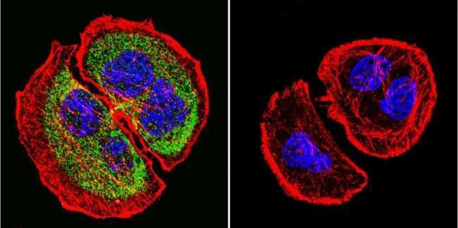
- Immunofluorescent analysis of PRC1 using PRC1 Monoclonal Antibody (16F2) (Product # MA1-846) shows staining in U251 Cells. PRC1 (green), F-Actin staining with Phalloidin (red) and nuclei with DAPI (blue) is shown. Cells were grown on chamber slides and fixed with formaldehyde prior to staining. Cells were probed without (control) or with an antibody recognizing PRC1 (Product # MA1-846) at a dilution of 1:20 over night at 4 °C, washed with PBS and incubated with a DyLight-488 conjugated secondary antibody (Product # 35552 for GAR, Product # 35503 for GAM). Images were taken at 60X magnification.
- Submitted by
- Invitrogen Antibodies (provider)
- Main image
- Experimental details
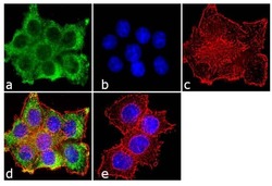
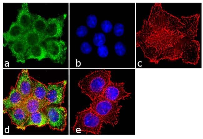
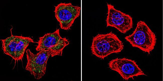
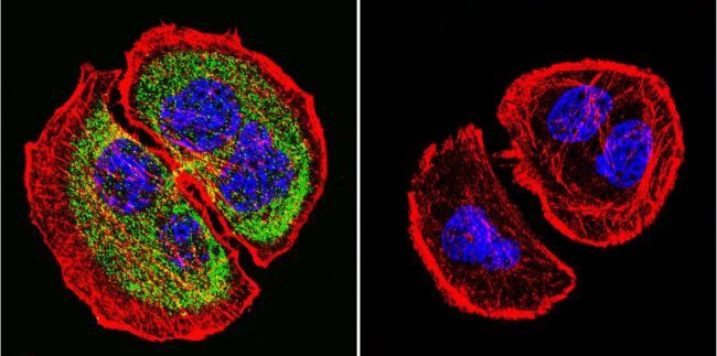
- Immunofluorescence analysis of PRC1 was done on 70% confluent log phase HeLa cells. The cells were fixed with 4% paraformaldehyde for 10 minutes, permeabilized with 0.1% Triton™ X-100 for 10 minutes, and blocked with 1% BSA for 1 hour at room temperature. The cells were labeled with PRC1 (16F2) Mouse Monoclonal Antibody (Product # MA1-846) at 2 µg/mL in 0.1% BSA and incubated for 3 hours at room temperature and then labeled with Goat anti-Mouse IgG (H+L) Superclonal™ Secondary Antibody, Alexa Fluor® 488 conjugate (Product # A28175) at a dilution of 1:2000 for 45 minutes at room temperature (Panel a: green). Nuclei (Panel b: blue) were stained with SlowFade® Gold Antifade Mountant with DAPI (Product # S36938). F-actin (Panel c: red) was stained with Alexa Fluor® 555 Rhodamine Phalloidin (Product # R415, 1:300). Panel d is a merged image showing cytoplasmic localization. Panel e is a no primary antibody control. The images were captured at 60X magnification.
- Submitted by
- Invitrogen Antibodies (provider)
- Main image

- Experimental details
- Immunofluorescence analysis of PRC1 was done on 70% confluent log phase HeLa cells. The cells were fixed with 4% paraformaldehyde for 10 minutes, permeabilized with 0.1% Triton™ X-100 for 10 minutes, and blocked with 1% BSA for 1 hour at room temperature. The cells were labeled with PRC1 (16F2) Mouse Monoclonal Antibody (Product # MA1-846) at 2 µg/mL in 0.1% BSA and incubated for 3 hours at room temperature and then labeled with Goat anti-Mouse IgG (H+L) Superclonal™ Secondary Antibody, Alexa Fluor® 488 conjugate (Product # A28175) at a dilution of 1:2000 for 45 minutes at room temperature (Panel a: green). Nuclei (Panel b: blue) were stained with SlowFade® Gold Antifade Mountant with DAPI (Product # S36938). F-actin (Panel c: red) was stained with Alexa Fluor® 555 Rhodamine Phalloidin (Product # R415, 1:300). Panel d is a merged image showing cytoplasmic localization. Panel e is a no primary antibody control. The images were captured at 60X magnification.

- Submitted by
- Invitrogen Antibodies (provider)
- Main image
- Experimental details
- Immunofluorescent analysis of PRC1 using PRC1 Monoclonal Antibody (16F2) (Product # MA1-846) shows staining in Hela Cells. PRC1 (green), F-Actin staining with Phalloidin (red) and nuclei with DAPI (blue) is shown. Cells were grown on chamber slides and fixed with formaldehyde prior to staining. Cells were probed without (control) or with an antibody recognizing PRC1 (Product # MA1-846) at a dilution of 1:100 over night at 4 °C, washed with PBS and incubated with a DyLight-488 conjugated secondary antibody (Product # 35552 for GAR, Product # 35503 for GAM). Images were taken at 60X magnification.
- Submitted by
- Invitrogen Antibodies (provider)
- Main image

- Experimental details
- Immunofluorescent analysis of PRC1 using PRC1 Monoclonal Antibody (16F2) (Product # MA1-846) shows staining in U251 Cells. PRC1 (green), F-Actin staining with Phalloidin (red) and nuclei with DAPI (blue) is shown. Cells were grown on chamber slides and fixed with formaldehyde prior to staining. Cells were probed without (control) or with an antibody recognizing PRC1 (Product # MA1-846) at a dilution of 1:20 over night at 4 °C, washed with PBS and incubated with a DyLight-488 conjugated secondary antibody (Product # 35552 for GAR, Product # 35503 for GAM). Images were taken at 60X magnification.
Supportive validation
- Submitted by
- Invitrogen Antibodies (provider)
- Main image
- Experimental details
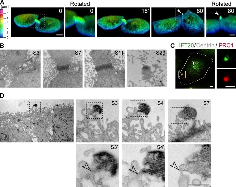
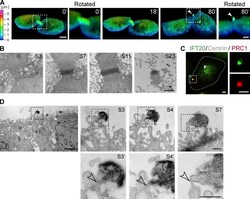
- Figure 2. The peripheral structure containing ciliary markers is a postmitotic midbody. (A) The images correspond to 3D reconstructions of cells expressing cherry-tubulin that were filmed during cell division. The images were pseudocolored based on height, using the color scale on the left, to highlight that the intercellular bridge forms at the top of the cells and that the postmitotic midbody remnant localizes after abscission at a peripheral position at the apical surface. The arrowhead points to the postmitotic midbody. Bar, 5 um. An enlargement of the boxed region at 0 and 80 min is also shown. Bars, 2 um. (B) Serial EM sections of the apical region of MDCK cells during cytokinesis. The sections are numbered S1 onwards from the lower section to the top. Note the progressive loss of microvilli at the top sections. Bar, 1 um. (C) Cells expressing dsRed-centrin were stained for IFT20 and PRC1. The enlargement of the boxed region shows the fluorescent signal for IFT20 and PRC1. The dashed line indicates the cell contour. The white and green arrowheads point to the centrosome and the peripheral structure, respectively. Bars, 2 um. (D) EM micrograph of a cell with a peripheral midbody remnant (left panel) and enlargements of different serial sections (S3, S4, and S7, right). Note that in some of the sections it appears that the remnant is connected to the rest of the cell by a thin tether (empty arrowhead) as shown in the enlargements of the boxed regions of sections S3, S4, a