Explore
 Validate
 Learn
Learn Western blot
Antibody data
- Antibody Data
- Antigen structure
- References [0]
- Comments [0]
- Validations
- Western blot [1]
- Immunocytochemistry [1]
- Immunohistochemistry [1]
- Flow cytometry [1]
Submit
Validation data
Reference
Comment
Report error
- Product number
- AP54517PU-N - Provider product page

- Provider
- Acris Antibodies GmbH
- Proper citation
- Acris Antibodies GmbH Cat#AP54517PU-N, RRID:AB_11142911
- Product name
- anti VPS52 (C-term)
- Antibody type
- Polyclonal
- Antigen
- KLH conjugated synthetic peptide between 613-641 amino acids from the C-terminal region of human VPS52
- Reactivity
- Human
- Host
- Rabbit
- Vial size
- 0.4 ml
- Concentration
- lot specific
No comments: Submit comment
Supportive validation
- Submitted by
- Acris Antibodies GmbH (provider)
- Main image

- Experimental details
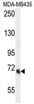
- VPS52 Antibody (C-term) (AP54517PU-N) western blot analysis in MDA-MB435 cell line lysates (35 µg/lane). This demonstrates the VPS52 antibody detected the VPS52 protein (arrow).
Supportive validation
- Submitted by
- Acris Antibodies GmbH (provider)
- Main image
- Experimental details
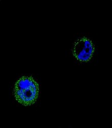
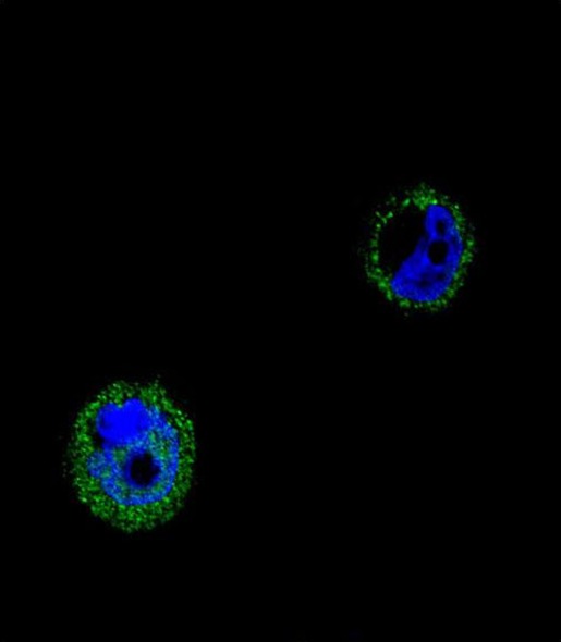
- Confocal immunofluorescent analysis of VPS52 Antibody (C-term) (AP54517PU-N) with MCF-7 cell followed by Alexa Fluor® 488-conjugated goat anti-rabbit lgG (green). DAPI was used to stain the cell nuclear (blue).
Supportive validation
- Submitted by
- Acris Antibodies GmbH (provider)
- Main image
- Experimental details
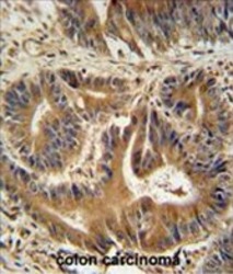
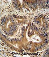
- VPS52 antibody (C-term) (AP54517PU-N) immunohistochemistry analysis in formalin fixed and paraffin embedded human colon carcinoma followed by peroxidase conjugation of the secondary antibody and DAB staining. This data demonstrates the use of the VPS52 antibody (C-term) for immunohistochemistry. Clinical relevance has not been evaluated.
Supportive validation
- Submitted by
- Acris Antibodies GmbH (provider)
- Main image
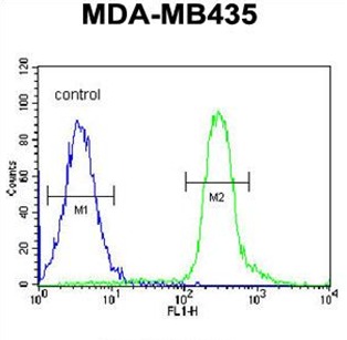
- Experimental details
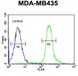
- VPS52 Antibody (C-term) (AP54517PU-N) flow cytometric analysis of MDA-MB435 cells (right histogram) compared to a negative control cell (left histogram). FITC-conjugated goat-anti-rabbit secondary antibodies were used for the analysis.