Explore
 Validate
 Learn
Learn Western blot
Antibody data
- Antibody Data
- Antigen structure
- References [1]
- Comments [0]
- Validations
- Western blot [1]
- Other assay [1]
Submit
Validation data
Reference
Comment
Report error
- Product number
- PA5-75980 - Provider product page

- Provider
- Invitrogen Antibodies
- Product name
- PMEPA1 Polyclonal Antibody
- Antibody type
- Polyclonal
- Antigen
- Synthetic peptide
- Description
- The antibody was affinity-purified from rabbit antiserum by affinity-chromatography using epitope-specific immunogen and the purity is > 100% (by SDS-PAGE).
- Reactivity
- Human, Mouse, Rat
- Host
- Rabbit
- Isotype
- IgG
- Vial size
- 100 µL
- Concentration
- 1 mg/mL
- Storage
- Store at 4°C short term. For long term storage, store at -20°C, avoiding freeze/thaw cycles.
Submitted references Hdac3 deletion in myeloid progenitor cells enhances bone healing in females and limits osteoclast fusion via Pmepa1.
Molstad DHH, Zars E, Norton A, Mansky KC, Westendorf JJ, Bradley EW
Scientific reports 2020 Dec 11;10(1):21804
Scientific reports 2020 Dec 11;10(1):21804
No comments: Submit comment
Supportive validation
- Submitted by
- Invitrogen Antibodies (provider)
- Main image

- Experimental details
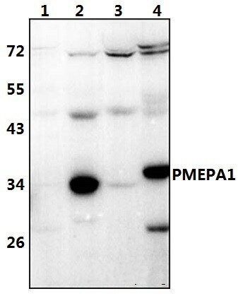
- Western blot analysis of PMEPA1 in Lane 1: HeLa whole cell lysate (40 µg), Lane 2: PC3 whole cell lysate (40 µg), Lane 3: the uterus tissue lysate of mouse (40 µg), Lane 4: the testis tissue lysate of rat (40 µg). Samples were incubated with PMEPA1 polyclonal antibody (Product # PA5-75980) at a dilution of 1:500.
Supportive validation
- Submitted by
- Invitrogen Antibodies (provider)
- Main image
- Experimental details
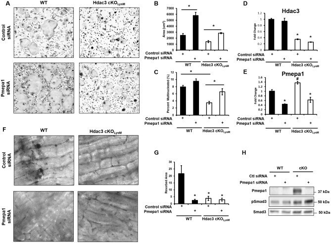
- Figure 7 Pmepa1 knock-down rescues Hdac3 deficiency. Bone marrow macrophages were collected from Hdac3 cKO LysM 6-week-old male or female mice or their control Cre + littermates and used to generate osteoclasts ex vivo. Shown are cultures from female mice. On day 4 cells were TRAP stained ( A ) and ( B ) osteoclast area and ( C ) percent multinucleated cells was determined. * p < 0.05. Cells were also seeded onto bone slices. On day 14, resorption pits were stained with Toluidine blue ( F ) and quantified ( G ). * p < 0.05 Expression levels of ( D ) Hdac3 and ( E ) Pmepa1 were evaluated by qPCR. * p < 0.05 Western blotting confirmed knockdown of Pmepa1 ( F ). Pit formation assays were also performed ( H ).