Explore
 Validate
 Learn
Learn Immunocytochemistry
Antibody data
- Antibody Data
- Antigen structure
- References [10]
- Comments [0]
- Validations
- Immunocytochemistry [1]
- Immunohistochemistry [1]
Submit
Validation data
Reference
Comment
Report error
- Product number
- HPA021057 - Provider product page

- Provider
- Atlas Antibodies
- Proper citation
- Atlas Antibodies Cat#HPA021057, RRID:AB_1848586
- Product name
- Anti-FBN1
- Antibody type
- Polyclonal
- Description
- Polyclonal Antibody against Human FBN1, Gene description: fibrillin 1, Alternative Gene Names: FBN, MASS, MFS1, OCTD, SGS, WMS, Validated applications: IHC, ICC, Uniprot ID: P35555, Storage: Store at +4°C for short term storage. Long time storage is recommended at -20°C.
- Reactivity
- Human
- Host
- Rabbit
- Conjugate
- Unconjugated
- Isotype
- IgG
- Vial size
- 100 µl
- Concentration
- 0.1 mg/ml
- Storage
- Store at +4°C for short term storage. Long time storage is recommended at -20°C.
- Handling
- The antibody solution should be gently mixed before use.
Submitted references A non-canonical role of ELN protects from cellular senescence by limiting iron-dependent regulation of gene expression
Transcriptomic alterations underlying metaplasia into specific metaplastic components in metaplastic breast carcinoma
A positively selected FBN1 missense variant reduces height in Peruvian individuals
Reversion‐inducing cysteine‐rich protein with Kazal motifs and MT1‐MMP promote the formation of robust fibrillin fibers
A proteome comparison between human fetal and mature renal extracellular matrix identifies EMILIN1 as a regulator of renal epithelial cell adhesion
ADAMTS-10 and -6 differentially regulate cell-cell junctions and focal adhesions
Network-based Survival Analysis Reveals Subnetwork Signatures for Predicting Outcomes of Ovarian Cancer Treatment
Epithelial-mesenchymal status influences how cells deposit fibrillin microfibrils
CD99 is a novel prognostic stromal marker in non‐small cell lung cancer
Czarnecka-Herok J, Zhu K, Flaman J, Goehrig D, Vernier M, Makulyte G, Lamboux A, Dragic H, Rhinn M, Médard J, Faury G, Bertolino P, Balter V, Debret R, Adnot S, Martin N, Bernard D
Redox Biology 2024;73
Redox Biology 2024;73
Michaud M, Mota L, Bakhtiari M, Thomas B, Tomeo J, Pilcher W, Contreras M, Ferran C, Bhasin S, Pradhan-Nabzdyk L, LoGerfo F, Liang P, Bhasin M
2024
2024
Transcriptomic alterations underlying metaplasia into specific metaplastic components in metaplastic breast carcinoma
Lien H, Hsu C, Lu Y, Chen T, Chen I, Li Y, Huang C, Cheng A, Lin C
Breast Cancer Research 2023;25(1)
Breast Cancer Research 2023;25(1)
A positively selected FBN1 missense variant reduces height in Peruvian individuals
Asgari S, Luo Y, Akbari A, Belbin G, Li X, Harris D, Selig M, Bartell E, Calderon R, Slowikowski K, Contreras C, Yataco R, Galea J, Jimenez J, Coit J, Farroñay C, Nazarian R, O’Connor T, Dietz H, Hirschhorn J, Guio H, Lecca L, Kenny E, Freeman E, Murray M, Raychaudhuri S
Nature 2020;582(7811):234-239
Nature 2020;582(7811):234-239
Reversion‐inducing cysteine‐rich protein with Kazal motifs and MT1‐MMP promote the formation of robust fibrillin fibers
Matsuzaki T, Keene D, Nishimoto E, Noda M
Journal of Cellular Physiology 2020;236(3):1980-1995
Journal of Cellular Physiology 2020;236(3):1980-1995
A proteome comparison between human fetal and mature renal extracellular matrix identifies EMILIN1 as a regulator of renal epithelial cell adhesion
Louzao-Martinez L, van Dijk C, Xu Y, Korn A, Bekker N, Brouwhuis R, Nicese M, Demmers J, Goumans M, Masereeuw R, Duncker D, Verhaar M, Cheng C
Matrix Biology Plus 2019;4
Matrix Biology Plus 2019;4
ADAMTS-10 and -6 differentially regulate cell-cell junctions and focal adhesions
Cain S, Mularczyk E, Singh M, Massam-Wu T, Kielty C
Scientific Reports 2016;6(1)
Scientific Reports 2016;6(1)
Network-based Survival Analysis Reveals Subnetwork Signatures for Predicting Outcomes of Ovarian Cancer Treatment
Miyano S, Zhang W, Ota T, Shridhar V, Chien J, Wu B, Kuang R
PLoS Computational Biology 2013;9(3):e1002975
PLoS Computational Biology 2013;9(3):e1002975
Epithelial-mesenchymal status influences how cells deposit fibrillin microfibrils
Baldwin A, Cain S, Lennon R, Godwin A, Merry C, Kielty C
Journal of Cell Science 2013
Journal of Cell Science 2013
CD99 is a novel prognostic stromal marker in non‐small cell lung cancer
Edlund K, Lindskog C, Saito A, Berglund A, Pontén F, Göransson‐Kultima H, Isaksson A, Jirström K, Planck M, Johansson L, Lambe M, Holmberg L, Nyberg F, Ekman S, Bergqvist M, Landelius P, Lamberg K, Botling J, Östman A, Micke P
International Journal of Cancer 2012;131(10):2264-2273
International Journal of Cancer 2012;131(10):2264-2273
No comments: Submit comment
Supportive validation
- Submitted by
- Atlas Antibodies (provider)
- Main image
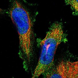
- Experimental details
- Immunofluorescent staining of human cell line U-251 MG shows localization to cytosol.
- Sample type
- Human

Supportive validation
- Submitted by
- Atlas Antibodies (provider)
- Enhanced method
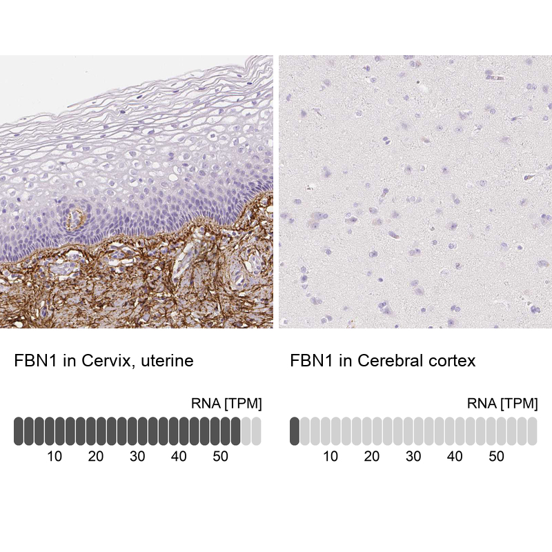
- Orthogonal validation
- Main image
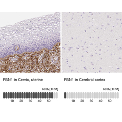
- Experimental details
- Immunohistochemistry analysis in human cervix, uterine and cerebral cortex tissues using HPA021057 antibody. Corresponding FBN1 RNA-seq data are presented for the same tissues.
- Sample type
- Human
- Protocol
- Protocol