Explore
 Validate
 Learn
Learn Western blot
 Immunocytochemistry
Antibody data
- Antibody Data
- Antigen structure
- References [24]
- Comments [0]
- Validations
- Immunocytochemistry [3]
- Other assay [1]
Submit
Validation data
Reference
Comment
Report error
- Product number
- MA5-12770 - Provider product page

- Provider
- Invitrogen Antibodies
- Product name
- Fibrillin 1 Monoclonal Antibody (11C1.3)
- Antibody type
- Monoclonal
- Antigen
- Other
- Description
- MA5-12770 targets Fibrillin-1 in ICC/IF, IHC (F), IM, and WB applications and shows reactivity with Bovine and Human samples. The MA5-12770 immunogen is microfibrils from zonular apparatus of bovine eye.
- Reactivity
- Human, Bovine
- Host
- Mouse
- Isotype
- IgG
- Antibody clone number
- 11C1.3
- Vial size
- 500 µL
- Concentration
- 0.2 mg/mL
- Storage
- 4° C
Submitted references Targeting feed-forward signaling of TGFβ/NOX4/DHFR/eNOS uncoupling/TGFβ axis with anti-TGFβ and folic acid attenuates formation of aortic aneurysms: Novel mechanisms and therapeutics.
Biochemical changes of the pericellular matrix and spatial chondrocyte organization-Two highly interconnected hallmarks of osteoarthritis.
Transcriptome analysis of skin fibroblasts with dominant negative COL3A1 mutations provides molecular insights into the etiopathology of vascular Ehlers-Danlos syndrome.
Results from in vitro and ex vivo skin aging models assessing the antiglycation and anti-elastase MMP-12 potential of glycylglycine oleamide.
GLUT10 deficiency leads to oxidative stress and non-canonical αvβ3 integrin-mediated TGFβ signalling associated with extracellular matrix disarray in arterial tortuosity syndrome skin fibroblasts.
Microfibril-associated glycoprotein-1 controls human ciliary zonule development in vitro.
Geographical ancestry is a key determinant of epidermal morphology and dermal composition.
Matrix metalloproteinase-2 degrades fibrillin-1 and fibrillin-2 of oxytalan fibers in the human eye and periodontal ligaments in vitro.
Latent transforming growth factor-β binding protein 2 negatively regulates coalescence of oxytalan fibers induced by stretching stress.
Tomato paste rich in lycopene protects against cutaneous photodamage in humans in vivo: a randomized controlled trial.
Dermal tissue and cellular expression of fibrillin-1 in diffuse cutaneous systemic sclerosis.
Cellular senescence of human mammary epithelial cells (HMEC) is associated with an altered MMP-7/HB-EGF signaling and increased formation of elastin-like structures.
Effects of a cosmetic 'anti-ageing' product improves photoaged skin [corrected].
A dermal equivalent developed from fibroblast culture alone: effect of EGF and insulin.
Increased expression of extracellular matrix proteins in rapid atrial pacing-induced atrial fibrillation.
Photoprotective and anti-skin-aging effects of eicosapentaenoic acid in human skin in vivo.
Induction of apoptosis and fibrillin 1 expression in human dermal endothelial cells by scleroderma sera containing anti-endothelial cell antibodies.
Sex steroids modulate human aortic smooth muscle cell matrix protein deposition and matrix metalloproteinase expression.
Ramipril reduces large-artery stiffness in peripheral arterial disease and promotes elastogenic remodeling in cell culture.
Heat modulation of tropoelastin, fibrillin-1, and matrix metalloproteinase-12 in human skin in vivo.
The effects of a novel synthetic retinoid, seletinoid G, on the expression of extracellular matrix proteins in aged human skin in vivo.
Expression of fibrillin-1 in focal nodular hyperplasia of the liver: a role in microcirculation adaptability.
Cell adhesion to fibrillin-1 molecules and microfibrils is mediated by alpha 5 beta 1 and alpha v beta 3 integrins.
Abnormal hepatic expression of fibrillin-1 in children with cholestasis.
Huang K, Wang Y, Siu KL, Zhang Y, Cai H
Redox biology 2021 Jan;38:101757
Redox biology 2021 Jan;38:101757
Biochemical changes of the pericellular matrix and spatial chondrocyte organization-Two highly interconnected hallmarks of osteoarthritis.
Danalache M, Erler AL, Wolfgart JM, Schwitalle M, Hofmann UK
Journal of orthopaedic research : official publication of the Orthopaedic Research Society 2020 Oct;38(10):2170-2180
Journal of orthopaedic research : official publication of the Orthopaedic Research Society 2020 Oct;38(10):2170-2180
Transcriptome analysis of skin fibroblasts with dominant negative COL3A1 mutations provides molecular insights into the etiopathology of vascular Ehlers-Danlos syndrome.
Chiarelli N, Carini G, Zoppi N, Ritelli M, Colombi M
PloS one 2018;13(1):e0191220
PloS one 2018;13(1):e0191220
Results from in vitro and ex vivo skin aging models assessing the antiglycation and anti-elastase MMP-12 potential of glycylglycine oleamide.
Bogdanowicz P, Haure MJ, Ceruti I, Bessou-Touya S, Castex-Rizzi N
Clinical, cosmetic and investigational dermatology 2016;9:143-50
Clinical, cosmetic and investigational dermatology 2016;9:143-50
GLUT10 deficiency leads to oxidative stress and non-canonical αvβ3 integrin-mediated TGFβ signalling associated with extracellular matrix disarray in arterial tortuosity syndrome skin fibroblasts.
Zoppi N, Chiarelli N, Cinquina V, Ritelli M, Colombi M
Human molecular genetics 2015 Dec 1;24(23):6769-87
Human molecular genetics 2015 Dec 1;24(23):6769-87
Microfibril-associated glycoprotein-1 controls human ciliary zonule development in vitro.
Fujita T, Tsuruga E, Yamanouchi K, Sawa Y, Ishikawa H
Acta histochemica et cytochemica 2014 Feb 27;47(1):11-7
Acta histochemica et cytochemica 2014 Feb 27;47(1):11-7
Geographical ancestry is a key determinant of epidermal morphology and dermal composition.
Langton AK, Sherratt MJ, Sellers WI, Griffiths CE, Watson RE
The British journal of dermatology 2014 Aug;171(2):274-82
The British journal of dermatology 2014 Aug;171(2):274-82
Matrix metalloproteinase-2 degrades fibrillin-1 and fibrillin-2 of oxytalan fibers in the human eye and periodontal ligaments in vitro.
Kawagoe M, Tsuruga E, Oka K, Sawa Y, Ishikawa H
Acta histochemica et cytochemica 2013 Oct 30;46(5):153-9
Acta histochemica et cytochemica 2013 Oct 30;46(5):153-9
Latent transforming growth factor-β binding protein 2 negatively regulates coalescence of oxytalan fibers induced by stretching stress.
Tsuruga E, Oka K, Hatakeyama Y, Isokawa K, Sawa Y
Connective tissue research 2012;53(6):521-7
Connective tissue research 2012;53(6):521-7
Tomato paste rich in lycopene protects against cutaneous photodamage in humans in vivo: a randomized controlled trial.
Rizwan M, Rodriguez-Blanco I, Harbottle A, Birch-Machin MA, Watson RE, Rhodes LE
The British journal of dermatology 2011 Jan;164(1):154-62
The British journal of dermatology 2011 Jan;164(1):154-62
Dermal tissue and cellular expression of fibrillin-1 in diffuse cutaneous systemic sclerosis.
Wipff J, Avouac J, Le Charpentier M, Varret M, Houtteman A, Ruiz B, Vacher-Lavenu MC, Kahan A, Boileau C, Allanore Y
Rheumatology (Oxford, England) 2010 Apr;49(4):657-61
Rheumatology (Oxford, England) 2010 Apr;49(4):657-61
Cellular senescence of human mammary epithelial cells (HMEC) is associated with an altered MMP-7/HB-EGF signaling and increased formation of elastin-like structures.
Bertram C, Hass R
Mechanisms of ageing and development 2009 Oct;130(10):657-69
Mechanisms of ageing and development 2009 Oct;130(10):657-69
Effects of a cosmetic 'anti-ageing' product improves photoaged skin [corrected].
Watson RE, Ogden S, Cotterell LF, Bowden JJ, Bastrilles JY, Long SP, Griffiths CE
The British journal of dermatology 2009 Aug;161(2):419-26
The British journal of dermatology 2009 Aug;161(2):419-26
A dermal equivalent developed from fibroblast culture alone: effect of EGF and insulin.
Lee DY, Yang JM, Park KH
Wound repair and regeneration : official publication of the Wound Healing Society [and] the European Tissue Repair Society 2007 Nov-Dec;15(6):936-9
Wound repair and regeneration : official publication of the Wound Healing Society [and] the European Tissue Repair Society 2007 Nov-Dec;15(6):936-9
Increased expression of extracellular matrix proteins in rapid atrial pacing-induced atrial fibrillation.
Lin CS, Lai LP, Lin JL, Sun YL, Hsu CW, Chen CL, Mao SJ, Huang SK
Heart rhythm 2007 Jul;4(7):938-49
Heart rhythm 2007 Jul;4(7):938-49
Photoprotective and anti-skin-aging effects of eicosapentaenoic acid in human skin in vivo.
Kim HH, Cho S, Lee S, Kim KH, Cho KH, Eun HC, Chung JH
Journal of lipid research 2006 May;47(5):921-30
Journal of lipid research 2006 May;47(5):921-30
Induction of apoptosis and fibrillin 1 expression in human dermal endothelial cells by scleroderma sera containing anti-endothelial cell antibodies.
Ahmed SS, Tan FK, Arnett FC, Jin L, Geng YJ
Arthritis and rheumatism 2006 Jul;54(7):2250-62
Arthritis and rheumatism 2006 Jul;54(7):2250-62
Sex steroids modulate human aortic smooth muscle cell matrix protein deposition and matrix metalloproteinase expression.
Natoli AK, Medley TL, Ahimastos AA, Drew BG, Thearle DJ, Dilley RJ, Kingwell BA
Hypertension (Dallas, Tex. : 1979) 2005 Nov;46(5):1129-34
Hypertension (Dallas, Tex. : 1979) 2005 Nov;46(5):1129-34
Ramipril reduces large-artery stiffness in peripheral arterial disease and promotes elastogenic remodeling in cell culture.
Ahimastos AA, Natoli AK, Lawler A, Blombery PA, Kingwell BA
Hypertension (Dallas, Tex. : 1979) 2005 Jun;45(6):1194-9
Hypertension (Dallas, Tex. : 1979) 2005 Jun;45(6):1194-9
Heat modulation of tropoelastin, fibrillin-1, and matrix metalloproteinase-12 in human skin in vivo.
Chen Z, Seo JY, Kim YK, Lee SR, Kim KH, Cho KH, Eun HC, Chung JH
The Journal of investigative dermatology 2005 Jan;124(1):70-8
The Journal of investigative dermatology 2005 Jan;124(1):70-8
The effects of a novel synthetic retinoid, seletinoid G, on the expression of extracellular matrix proteins in aged human skin in vivo.
Kim MS, Lee S, Rho HS, Kim DH, Chang IS, Chung JH
Clinica chimica acta; international journal of clinical chemistry 2005 Dec;362(1-2):161-9
Clinica chimica acta; international journal of clinical chemistry 2005 Dec;362(1-2):161-9
Expression of fibrillin-1 in focal nodular hyperplasia of the liver: a role in microcirculation adaptability.
Lepreux S, Desmouliere A, Rosenbaum J, Balabaud C, Bioulac-Sage P
Comparative hepatology 2004 Jan 14;3 Suppl 1(Suppl 1):S57
Comparative hepatology 2004 Jan 14;3 Suppl 1(Suppl 1):S57
Cell adhesion to fibrillin-1 molecules and microfibrils is mediated by alpha 5 beta 1 and alpha v beta 3 integrins.
Bax DV, Bernard SE, Lomas A, Morgan A, Humphries J, Shuttleworth CA, Humphries MJ, Kielty CM
The Journal of biological chemistry 2003 Sep 5;278(36):34605-16
The Journal of biological chemistry 2003 Sep 5;278(36):34605-16
Abnormal hepatic expression of fibrillin-1 in children with cholestasis.
Lamireau T, Dubuisson L, Lepreux S, Bioulac-Sage P, Fabre M, Rosenbaum J, Desmoulière A
The American journal of surgical pathology 2002 May;26(5):637-46
The American journal of surgical pathology 2002 May;26(5):637-46
No comments: Submit comment
Supportive validation
- Submitted by
- Invitrogen Antibodies (provider)
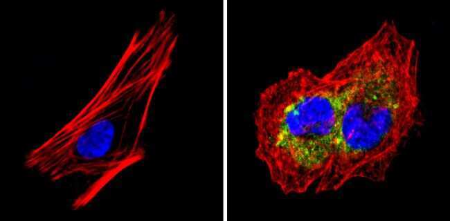
- Main image
- Experimental details
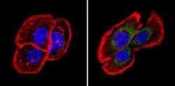
- Immunofluorescent analysis of Fibrillin-1 (green) showing staining in the cytoplasm of A431 cells (right) compared to a negative control without primary antibody (left). Formalin-fixed cells were permeabilized with 0.1% Triton X-100 in TBS for 5-10 minutes and blocked with 3% BSA-PBS for 30 minutes at room temperature. Cells were probed with a Fibrillin-1 monoclonal antibody (Product # MA5-12770) in 3% BSA-PBS at a dilution of 1:20 and incubated overnight at 4 ºC in a humidified chamber. Cells were washed with PBST and incubated with a DyLight-conjugated secondary antibody in PBS at room temperature in the dark. F-actin (red) was stained with a fluorescent red phalloidin and nuclei (blue) were stained with Hoechst or DAPI. Images were taken at a magnification of 60x.
- Submitted by
- Invitrogen Antibodies (provider)
- Main image
- Experimental details
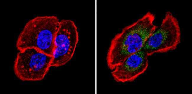
- Immunofluorescent analysis of Fibrillin-1 (green) showing staining in the cytoplasm of SH-SY5Y cells (right) compared to a negative control without primary antibody (left). Formalin-fixed cells were permeabilized with 0.1% Triton X-100 in TBS for 5-10 minutes and blocked with 3% BSA-PBS for 30 minutes at room temperature. Cells were probed with a Fibrillin-1 monoclonal antibody (Product # MA5-12770) in 3% BSA-PBS at a dilution of 1:100 and incubated overnight at 4 ºC in a humidified chamber. Cells were washed with PBST and incubated with a DyLight-conjugated secondary antibody in PBS at room temperature in the dark. F-actin (red) was stained with a fluorescent red phalloidin and nuclei (blue) were stained with Hoechst or DAPI. Images were taken at a magnification of 60x.
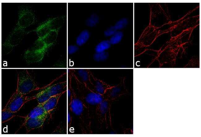
- Submitted by
- Invitrogen Antibodies (provider)
- Main image

- Experimental details
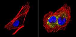
- Immunofluorescence analysis of Fibrillin-1 was performed using 70% confluent log phase SH-SY5Y cells. The cells were fixed with 4% paraformaldehyde for 10 minutes, permeabilized with 0.1% Triton™ X-100 for 10 minutes, and blocked with 1% BSA for 1 hour at room temperature. The cells were labeled with Fibrillin-1 (11C1.3) Mouse Monoclonal Antibody (Product # MA5-12770) at 2µg/mL in 0.1% BSA and incubated for 3 hours at room temperature and then labeled with Goat anti-Mouse IgG (H+L) Superclonal™ Secondary Antibody, Alexa Fluor® 488 conjugate (Product # A28175) at a dilution of 1:2000 for 45 minutes at room temperature (Panel a: green). Nuclei (Panel b: blue) were stained with SlowFade® Gold Antifade Mountant with DAPI (Product # S36938). F-actin (Panel c: red) was stained with Rhodamine Phalloidin (Product # R415, 1:300). Panel d represents the merged image showing cytoplasmic localization. Panel e shows the no primary antibody control. The images were captured at 60X magnification.
Supportive validation
- Submitted by
- Invitrogen Antibodies (provider)
- Main image
- Experimental details
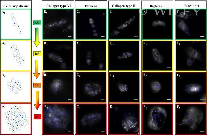
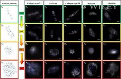
- 1 Figure Top-down view of immunohistochemical analysis of the pericellular matrix (PCM). Images are displayed in their physiopathological order of cellular spatial organization. A 1 /B 1 /C 1 /D 1 /E 1 /F 1 , single strings (enframed in green); A 2 /B 2 /C 2 /D 2 /E 2 /F 2 , double strings (enframed in yellow); A 3 /B 3 /C 3 /D 3 /E 3 /E 3 , small clusters (enframed in orange); A 4 /B 4 /C 4 /D 4 /E 4 /F 4 , big clusters (enframed in red). Immunolabelled PCM (depicted in white) for collagen type VI (B 1 -B 4 ), perlecan (C 1 -C 4 ), collagen type III (D 1 -D 4 ), biglycan (E 1 -E 4 ), and fibrillin-1 (F 1 -F 2 ). From single strings to big clusters, a decrease in signal intensity and pericellular definition can be observed for collagen type VI (B 1 -B 4 ), perlecan (C 1 -C 4 ), and biglycan (E 1 -E 4 ). Signal intensity remains constant, however, for collagen type III (D 1 -D 4 ) and fibrillin-1 (F 1 -F 4 ). Twenty-fold magnification; scale bars represent 20 um. Nuclei are stained in blue (DAPI), the stained PCM components are visualized in white and the cell membrane is depicted by black circles. B 1 -B 4 and C 1 -C 4 adapted from Danalache et al. 39 BC, big clusters; DAPI, 4',6-diamidino-2-phenylindole; SC, small clusters [Color figure can be viewed at wileyonlinelibrary.com ]