Explore
 Validate
 Learn
Learn Western blot
 Immunohistochemistry
Antibody data
- Antibody Data
- Antigen structure
- References [10]
- Comments [0]
- Validations
- Western blot [2]
- Immunocytochemistry [2]
Submit
Validation data
Reference
Comment
Report error
- Product number
- HPA003037 - Provider product page

- Provider
- Atlas Antibodies
- Proper citation
- Atlas Antibodies Cat#HPA003037, RRID:AB_1080329
- Product name
- Anti-TOMM22
- Antibody type
- Polyclonal
- Description
- Polyclonal Antibody against Human TOMM22, Gene description: translocase of outer mitochondrial membrane 22 homolog (yeast), Alternative Gene Names: TOM22, Validated applications: WB, ICC, IHC, Uniprot ID: Q9NS69, Storage: Store at +4°C for short term storage. Long time storage is recommended at -20°C.
- Reactivity
- Human
- Host
- Rabbit
- Conjugate
- Unconjugated
- Isotype
- IgG
- Vial size
- 100 µl
- Concentration
- 0.2 mg/ml
- Storage
- Store at +4°C for short term storage. Long time storage is recommended at -20°C.
- Handling
- The antibody solution should be gently mixed before use.
Submitted references In vivo CRISPR screening directly targeting testicular cells
Culture of Bovine Aortic Endothelial Cells in Galactose Media Enhances Mitochondrial Plasticity and Changes Redox Sensing, Altering Nrf2 and FOXO3 Levels.
Evaluation of Mitochondrial Function in Blood Samples Shows Distinct Patterns in Subjects with Thyroid Carcinoma from Those with Hyperplasia
Functional Analysis of GSTK1 in Peroxisomal Redox Homeostasis in HEK-293 Cells.
Defining the interactome of the human mitochondrial ribosome identifies SMIM4 and TMEM223 as respiratory chain assembly factors
Loss of MTX2 causes mandibuloacral dysplasia and links mitochondrial dysfunction to altered nuclear morphology
Two different pathogenic mechanisms, dying-back axonal neuropathy and pancreatic senescence, are present in the YG8R mouse model of Friedreich’s ataxia
Global variability in gene expression and alternative splicing is modulated by mitochondrial content
Global variability in gene expression and alternative splicing is modulated by mitochondrial content
Skeletal muscle stem cells adopt a dormant cell state post mortem and retain regenerative capacity
Noguchi Y, Onodera Y, Miyamoto T, Maruoka M, Kosako H, Suzuki J
Cell Genomics 2024;4(3):100510
Cell Genomics 2024;4(3):100510
Culture of Bovine Aortic Endothelial Cells in Galactose Media Enhances Mitochondrial Plasticity and Changes Redox Sensing, Altering Nrf2 and FOXO3 Levels.
Galant LS, Doblado L, Radi R, de Bem AF, Monsalve M
Antioxidants (Basel, Switzerland) 2024 Jul 20;13(7)
Antioxidants (Basel, Switzerland) 2024 Jul 20;13(7)
Evaluation of Mitochondrial Function in Blood Samples Shows Distinct Patterns in Subjects with Thyroid Carcinoma from Those with Hyperplasia
Bernal-Tirapo J, Bayo Jiménez M, Yuste-García P, Cordova I, Peñas A, García-Borda F, Quintela C, Prieto I, Sánchez-Ramos C, Ferrero-Herrero E, Monsalve M
International Journal of Molecular Sciences 2023;24(7):6453
International Journal of Molecular Sciences 2023;24(7):6453
Functional Analysis of GSTK1 in Peroxisomal Redox Homeostasis in HEK-293 Cells.
Costa CF, Lismont C, Chornyi S, Li H, Hussein MAF, Waterham HR, Fransen M
Antioxidants (Basel, Switzerland) 2023 Jun 7;12(6)
Antioxidants (Basel, Switzerland) 2023 Jun 7;12(6)
Defining the interactome of the human mitochondrial ribosome identifies SMIM4 and TMEM223 as respiratory chain assembly factors
Poerschke S, Dennerlein S, Oeljeklaus S, Wang C, Richter-Dennerlein R, Sattmann J, Bauermeister D, Hanitsch E, Stoldt S, Langer T, Jakobs S, Warscheid B, Rehling P
eLife 2021;10
eLife 2021;10
Loss of MTX2 causes mandibuloacral dysplasia and links mitochondrial dysfunction to altered nuclear morphology
Elouej S, Harhouri K, Le Mao M, Baujat G, Nampoothiri S, Kayserili H, Menabawy N, Selim L, Paneque A, Kubisch C, Lessel D, Rubinsztajn R, Charar C, Bartoli C, Airault C, Deleuze J, Rötig A, Bauer P, Pereira C, Loh A, Escande-Beillard N, Muchir A, Martino L, Gruenbaum Y, Lee S, Manivet P, Lenaers G, Reversade B, Lévy N, De Sandre-Giovannoli A
Nature Communications 2020;11(1)
Nature Communications 2020;11(1)
Two different pathogenic mechanisms, dying-back axonal neuropathy and pancreatic senescence, are present in the YG8R mouse model of Friedreich’s ataxia
Mollá B, Riveiro F, Bolinches-Amorós A, Muñoz-Lasso D, Palau F, González-Cabo P
Disease Models & Mechanisms 2016;9(6):647-657
Disease Models & Mechanisms 2016;9(6):647-657
Global variability in gene expression and alternative splicing is modulated by mitochondrial content
Guantes R, Rastrojo A, Neves R, Lima A, Aguado B, Iborra F
Genome Research 2015;25(5):633-644
Genome Research 2015;25(5):633-644
Global variability in gene expression and alternative splicing is modulated by mitochondrial content
Guantes R, Rastrojo A, Neves R, Lima A, Aguado B, Iborra F
Genome Research 2015 May;25(5):633-644
Genome Research 2015 May;25(5):633-644
Skeletal muscle stem cells adopt a dormant cell state post mortem and retain regenerative capacity
Latil M, Rocheteau P, Châtre L, Sanulli S, Mémet S, Ricchetti M, Tajbakhsh S, Chrétien F
Nature Communications 2012;3(1)
Nature Communications 2012;3(1)
No comments: Submit comment
Enhanced validation
Enhanced validation
- Submitted by
- klas2
- Enhanced method
- Genetic validation
- Main image
- Experimental details
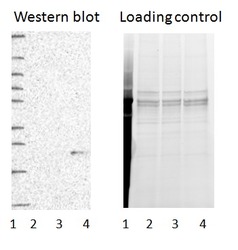
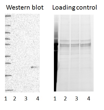
- Western blot of cell lysate from U-2 OS cells transfected with either siRNA targeting TOMM22 or control siRNA. Lane 1: Marker (250, 130, 95, 72, 55, 36, 28, 17, 10) Lane 2: Cell lysate from U-2OS cells transfected with siRNA targeting TOMM22 Lane 3: N/A Lane 4: Cell lysate from U-2OS cells transfected with control siRNA Right image, lane 1-4: loading control
- Sample type
- U-2 OS
- Primary Ab dilution
- 1:311
- Conjugate
- Horseradish Peroxidase
- Secondary Ab
- Secondary Ab
- Secondary Ab dilution
- 1:3000
- Knockdown/Genetic Approaches Application
- Western blot
Enhanced validation
- Submitted by
- Atlas Antibodies (provider)
- Enhanced method
- Genetic validation
- Main image

- Experimental details
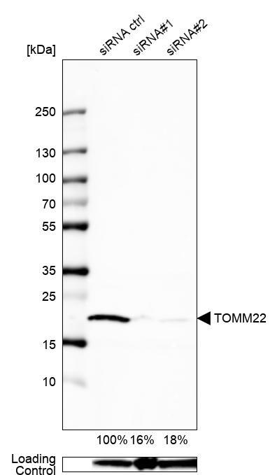
- Western blot analysis in U2OS cells transfected with control siRNA, target specific siRNA probe #1 and #2, using Anti-TOMM22 antibody. Remaining relative intensity is presented. Loading control: Anti-GAPDH.
- Sample type
- Human
- Protocol
- Protocol
Enhanced validation
Supportive validation
- Submitted by
- 55af80e3e0991
- Enhanced method
- Genetic validation
- Main image
- Experimental details
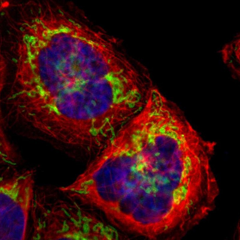
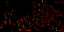
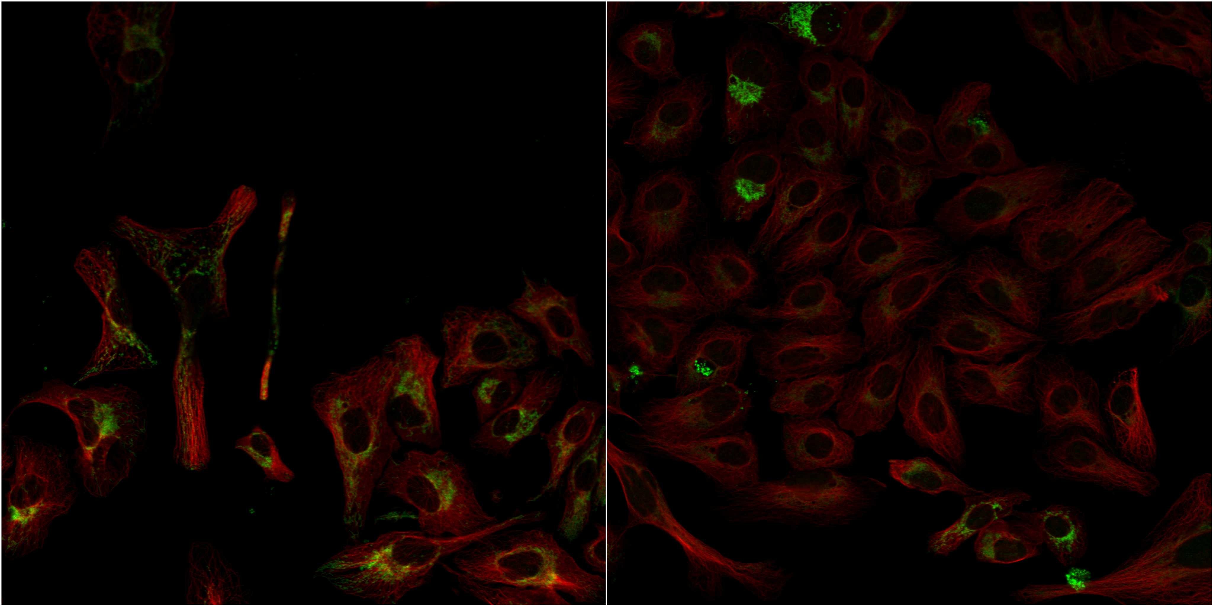
- Confocal images of immunofluorescently stained human U-2 OS cells.The protein TOMM22 is shown in green and the microtubules in red. The image to the left show cells transfected with control siRNA and the image to the right show cells where TOMM22 has been downregulated with specific siRNA.
- Sample type
- U-2 OS cells
- Primary Ab dilution
- 1:141
- Secondary Ab
- Secondary Ab
- Secondary Ab dilution
- 1:800
- Knockdown/Genetic Approaches Application
- Immunocytochemistry
Supportive validation
- Submitted by
- Atlas Antibodies (provider)
- Main image
- Experimental details
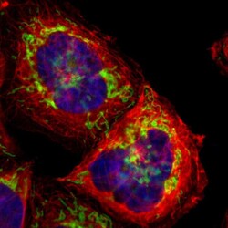
- Immunofluorescent staining of human cell line A-431 shows localization to mitochondria.
- Sample type
- Human