 Explore
 Validate
 Learn
LearnMA5-23767
antibody from Invitrogen Antibodies
Targeting: CCL26
eotaxin-3, IMAC, MIP-4a, MIP-4alpha, SCYA26, TSC-1
 Western blot
 ELISA
ELISA Other assay
Antibody data
- Antibody Data
- Antigen structure
- References [1]
- Comments [0]
- Validations
- Other assay [6]
Submit
Validation data
Reference
Comment
Report error
- Product number
- MA5-23767 - Provider product page

- Provider
- Invitrogen Antibodies
- Product name
- Eotaxin 3 Monoclonal Antibody (115002)
- Antibody type
- Monoclonal
- Antigen
- Recombinant full-length protein
- Description
- This antibody does not cross-react with recombinant human (rh) CCL11/Eotaxin, recombinant mouse (rm) CCL11/Eotaxin, rhCCL24/Eotaxin-2, rmCCL24/Eotaxin-2, rhCCL3, rhCCL4, rmCCL3, rmCCL4, rhCCL5, rhCCL13, or rhCCL14. Reconstitute at 0.5 mg/mL in sterile PBS. Endoxin level is
- Reactivity
- Human
- Host
- Mouse
- Isotype
- IgG
- Antibody clone number
- 115002
- Vial size
- 500 μg
- Concentration
- 0.5 mg/mL
- Storage
- -20°C, Avoid Freeze/Thaw Cycles
Submitted references Analysis of the signal cross talk via CCL26 in the tumor microenvironment in osteosarcoma.
Kawano M, Iwasaki T, Itonaga I, Kubota Y, Tanaka K, Tsumura H
Scientific reports 2021 Sep 13;11(1):18099
Scientific reports 2021 Sep 13;11(1):18099
No comments: Submit comment
Supportive validation
- Submitted by
- Invitrogen Antibodies (provider)
- Main image

- Experimental details
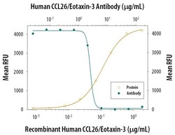
- Neutralization of Eotaxin 3 in BaF3 mouse pro‚B cell line transfected with mouse CCR3. Samples were incubated in Eotaxin 3 monoclonal antibody (Product # MA5-23858). Recombinant Human CCL26/Eotaxin‚3 chemoattracts the BaF3 mouse pro‚B cell line transfected with mouse CCR3 in a dose-dependent manner (orange line). The amount of cells that migrated through to the lower chemotaxis chamber was measured by Resazurin. Chemotaxis elicited by Recombinant Human CCL26/Eotaxin‚3 (1 µg/mL) is neutralized (green line) by increasing concentrations of Human CCL26/Eotaxin‚3 Monoclonal Antibody. The ND50 is typically 2-8 µg/mL.

- Submitted by
- Invitrogen Antibodies (provider)
- Main image
- Experimental details
- Neutralization of Eotaxin 3 in BaF3 mouse pro‚B cell line transfected with mouse CCR3. Samples were incubated in Eotaxin 3 monoclonal antibody (Product # MA5-23858). Recombinant Human CCL26/Eotaxin‚3 chemoattracts the BaF3 mouse pro‚B cell line transfected with mouse CCR3 in a dose-dependent manner (orange line). The amount of cells that migrated through to the lower chemotaxis chamber was measured by Resazurin. Chemotaxis elicited by Recombinant Human CCL26/Eotaxin‚3 (1 µg/mL) is neutralized (green line) by increasing concentrations of Human CCL26/Eotaxin‚3 Monoclonal Antibody. The ND50 is typically 2-8 µg/mL.
- Submitted by
- Invitrogen Antibodies (provider)
- Main image
- Experimental details
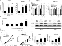
- Figure 2 Changes in CCL26 expression and cell growth in MG63 and hMSCs induced by the co-culture condition and rCCL26 administration. ( A ) Changes in CCL26 mRNA expression in MG63 and hMSCs were assessed by qRT-PCR. The co-culture and rCCL26 addition significantly increased the expression of mRNA of CCL26. ( B ) rCCL26 was administered to mono-cultured MG63. There was a significant increase in cell growth with rCCL26 at 10 ng/ml. (*) p < 0.05, (**) p < 0.01. ( C ) Changes in cell proliferation induced by Co-cultured condition and recombinant CCL26 administration in MG63 and hMSCs. ( D ) Changes in the degree of cell proliferation in the untreated, co-culture, and recombinant groups were analyzed by performing cell proliferation assay via BrdU incorporation. ( E ) Changes inCCL26 protein expression of intra-cellular hMSCs and MG63 were assessed by western blot analysis. The co-culture and addition of rCCL26 increased the expression ofCCL26 protein in MG63 and hMSCs. ( F ) The quantification of western blot analysis. Data represents represent the mean +- SD of three independent experiments. p < 0.05 was considered to indicate significance: (*) p < 0.05, (**) p < 0.01.

- Submitted by
- Invitrogen Antibodies (provider)
- Main image
- Experimental details
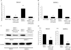
- Figure 3 Effects of neutralizing anti-CCL26 Ab on CCL26 expression in mono-cultured and co-cultured MG63 and hMSCs. ( A ) Changes inCCL26 expression in MG63 were assessed by qRT-PCR. The addition of antiCCL26 Ab to MG63 decreased the expression ofCCL26 mRNA. ( B ) Changes inCCL26 expression in hMSCs were assessed by qRT-PCR. The addition of anti-CCL26 Ab to hMSCs decreased the expression ofCCL26 mRNA. ( C ) Changes inCCL26 protein expression in hMSCs and MG63 were assessed by western blot analysis. The addition of anti-CCL26 Ab to MG63 and hMSCs decreased the expression ofCCL26 protein in these cells. ( D ) The quantification of western blot analysis. Data represents represent the mean +- SD of three independent experiments. p < 0.05 was considered to indicate significance: (*) p < 0.05, (**) p < 0.01.

- Submitted by
- Invitrogen Antibodies (provider)
- Main image

- Experimental details
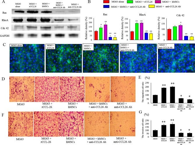
- Figure 4 Effects of co-culture condition and administration of rCCL26 or anti-CCL26 Ab on motility of MG63. ( A ) Changes in protein expression of factors related to cell motility. ( B ) The quantification of western blot analysis. Data represents represent the mean +- SD of three independent experiments. ( C ) The influence of co-culture and rCCL26 or anti-CCL26 Ab on actin fiber morphology was evaluated using immunofluorescent imaging. Original magnification, x 400; Scale bars: 50 mum. ( D ) The cell migration of MG63 was assessed in each group at 24 h after the challenge with or without rCCL26 and neutralizing anti-CCL26 Ab. ( E ) The amount of MG63 cells that crossed the membrane was measured. A significant decrease in motility was found in the group given anti-CCL26 Ab. ( F ) The cell invasion in MG63 was assessed in each group after 24 h. ( G ) The amount of the cells of which the membrane with Matrigel was crossed by MG63 was measured. Decreased migration ability was found in the group administered anti-CCL26 Ab. p < 0.05 was considered to indicate significance: (*) p < 0.05, (**) p < 0.01.
- Submitted by
- Invitrogen Antibodies (provider)
- Main image

- Experimental details
- Neutralization antibody testing demonstrates the specificity of an antibody through a correlation between antibody binding and the activity of the target. Neutralization of Eotaxin3 is shown by the decrease in fluorescence (Chemotaxis measured by Resazurin) with increasing concentrations of Eotaxin3 Antibody (Product # MA5-23767).
