Explore
 Validate
 Learn
Learn Western blot
 Immunocytochemistry
Antibody data
- Antibody Data
- Antigen structure
- References [6]
- Comments [0]
- Validations
- Immunocytochemistry [1]
- Immunohistochemistry [1]
Submit
Validation data
Reference
Comment
Report error
- Product number
- HPA013998 - Provider product page

- Provider
- Atlas Antibodies
- Proper citation
- Atlas Antibodies Cat#HPA013998, RRID:AB_1848016
- Product name
- Anti-NECAB2
- Antibody type
- Polyclonal
- Description
- Polyclonal Antibody against Human NECAB2, Gene description: N-terminal EF-hand calcium binding protein 2, Alternative Gene Names: EFCBP2, Validated applications: WB, IHC, ICC, Uniprot ID: Q7Z6G3, Storage: Store at +4°C for short term storage. Long time storage is recommended at -20°C.
- Reactivity
- Human, Mouse
- Host
- Rabbit
- Conjugate
- Unconjugated
- Isotype
- IgG
- Vial size
- 100 µl
- Concentration
- 0.3 mg/ml
- Storage
- Store at +4°C for short term storage. Long time storage is recommended at -20°C.
- Handling
- The antibody solution should be gently mixed before use.
Submitted references NECAB1-3, parvalbumin, calbindin, and calretinin in the hippocampus of the European mole.
Postnatal Developmental Expression Profile Classifies the Indusium Griseum as a Distinct Subfield of the Hippocampal Formation
NECAB1 and NECAB2 are Prevalent Calcium-Binding Proteins of CB1/CCK-Positive GABAergic Interneurons.
In vivo survival and differentiation of Friedreich ataxia iPSC-derived sensory neurons transplanted in the adult dorsal root ganglia.
Phenotypic and Functional Characterization of Peripheral Sensory Neurons derived from Human Embryonic Stem Cells
Neuronal calcium-binding proteins 1/2 localize to dorsal root ganglia and excitatory spinal neurons and are regulated by nerve injury
Maliković J, Amrein I, Vinciguerra L, Wolfer DP, Slomianka L
Frontiers in neuroanatomy 2024;18:1452722
Frontiers in neuroanatomy 2024;18:1452722
Postnatal Developmental Expression Profile Classifies the Indusium Griseum as a Distinct Subfield of the Hippocampal Formation
Sanders M, Petrasch-Parwez E, Habbes H, Düring M, Förster E
Frontiers in Cell and Developmental Biology 2021;8
Frontiers in Cell and Developmental Biology 2021;8
NECAB1 and NECAB2 are Prevalent Calcium-Binding Proteins of CB1/CCK-Positive GABAergic Interneurons.
Miczán V, Kelemen K, Glavinics JR, László ZI, Barti B, Kenesei K, Kisfali M, Katona I
Cerebral cortex (New York, N.Y. : 1991) 2021 Feb 5;31(3):1786-1806
Cerebral cortex (New York, N.Y. : 1991) 2021 Feb 5;31(3):1786-1806
In vivo survival and differentiation of Friedreich ataxia iPSC-derived sensory neurons transplanted in the adult dorsal root ganglia.
Viventi S, Frausin S, Howden SE, Lim SY, Finol-Urdaneta RK, McArthur JR, Abu-Bonsrah KD, Ng W, Ivanusic J, Thompson L, Dottori M
Stem cells translational medicine 2021 Aug;10(8):1157-1169
Stem cells translational medicine 2021 Aug;10(8):1157-1169
Phenotypic and Functional Characterization of Peripheral Sensory Neurons derived from Human Embryonic Stem Cells
Alshawaf A, Viventi S, Qiu W, D’Abaco G, Nayagam B, Erlichster M, Chana G, Everall I, Ivanusic J, Skafidas E, Dottori M
Scientific Reports 2018;8(1)
Scientific Reports 2018;8(1)
Neuronal calcium-binding proteins 1/2 localize to dorsal root ganglia and excitatory spinal neurons and are regulated by nerve injury
Zhang M, Tortoriello G, Hsueh B, Tomer R, Ye L, Mitsios N, Borgius L, Grant G, Kiehn O, Watanabe M, Uhlén M, Mulder J, Deisseroth K, Harkany T, Hökfelt T
Proceedings of the National Academy of Sciences 2014;111(12)
Proceedings of the National Academy of Sciences 2014;111(12)
No comments: Submit comment
Supportive validation
- Submitted by
- Atlas Antibodies (provider)
- Main image

- Experimental details
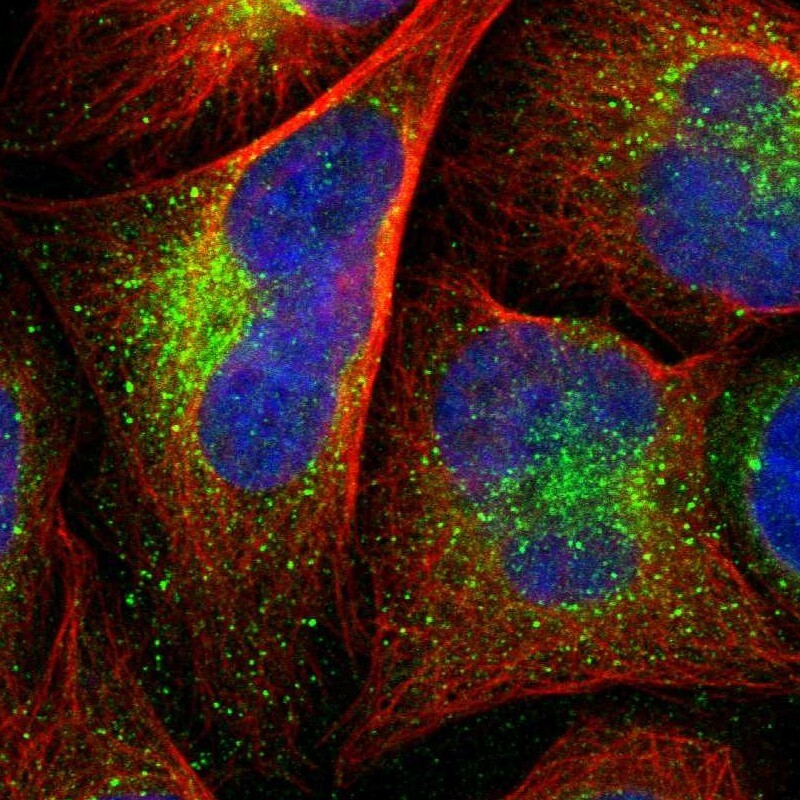
- Immunofluorescent staining of human cell line U-2 OS shows localization to vesicles.
- Sample type
- Human
Supportive validation
- Submitted by
- Atlas Antibodies (provider)
- Enhanced method
- Orthogonal validation
- Main image
- Experimental details
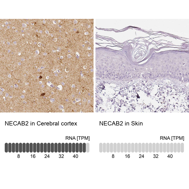
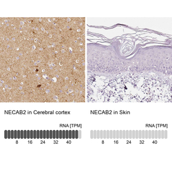
- Immunohistochemistry analysis in human cerebral cortex and skin tissues using HPA013998 antibody. Corresponding NECAB2 RNA-seq data are presented for the same tissues.
- Sample type
- Human
- Protocol
- Protocol