Explore
 Validate
 Learn
Learn Western blot
Antibody data
- Antibody Data
- Antigen structure
- References [0]
- Comments [0]
- Validations
- Western blot [2]
- ELISA [1]
- Immunohistochemistry [1]
Submit
Validation data
Reference
Comment
Report error
- Product number
- MAB97203-100 - Provider product page

- Provider
- R&D Systems
- Product name
- Human/Mouse/Rat Myoglobin Antibody
- Antibody type
- Monoclonal
- Description
- Protein A or G purified from cell culture supernatant. Detects human Myoglobin in direct ELISAs. Detects human, mouse, and rat Myoglabin in Western blots.
- Reactivity
- Human, Mouse, Rat
- Host
- Rabbit
- Conjugate
- Unconjugated
- Antigen sequence
P02144- Isotype
- IgG
- Antibody clone number
- 2269B
- Vial size
- 100 ug
- Storage
- Use a manual defrost freezer and avoid repeated freeze-thaw cycles. 12 months from date of receipt, -20 to -70 °C as supplied. 1 month, 2 to 8 °C under sterile conditions after reconstitution. 6 months, -20 to -70 °C under sterile conditions after reconstitution.
No comments: Submit comment
Supportive validation
- Submitted by
- R&D Systems (provider)
- Main image
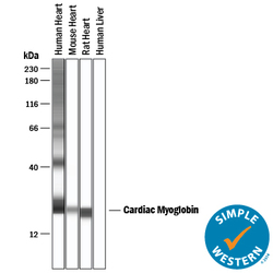
- Experimental details
- Detection of Human, Mouse, and Rat Myoglobin by Simple WesternTM. Simple Western lane view shows lysates of human heart tissue, mouse heart tissue, rat heart tissue, and human liver tissue, loaded at 0.2 mg/mL. A specific band was detected for Myoglobin at approximately 21-24 kDa (as indicated) using 10 µg/mL of Rabbit Anti-Human Myoglobin Monoclonal Antibody (Catalog # MAB97203) . This experiment was conducted under reducing conditions and using the 12-230 kDa separation system.

- Submitted by
- R&D Systems (provider)
- Main image
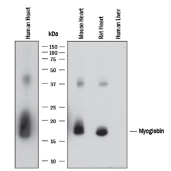
- Experimental details
- Detection of Human, Mouse, and Rat Myoglobin by Western Blot. Western blot shows lysates of human heart tissue, mouse heart tissue, rat heart tissue, and human liver tissue. PVDF membrane was probed with 0.5 µg/mL of Rabbit Anti-Human Myoglobin Monoclonal Antibody (Catalog # MAB97203) followed by HRP-conjugated Anti-Rabbit IgG Secondary Antibody (Catalog # HAF008). A specific band was detected for Myoglobin at approximately 18 kDa (as indicated). This experiment was conducted under reducing conditions and using Immunoblot Buffer Group 1.

Supportive validation
- Submitted by
- R&D Systems (provider)
- Main image
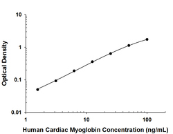
- Experimental details
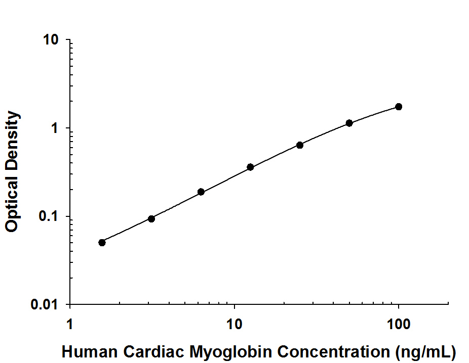
- Human Myoglobin ELISA Standard Curve. Recombinant Human Myoglobin protein was serially diluted 2-fold and captured by Rabbit Anti-Human Myoglobin Monoclonal Antibody (Catalog # MAB97204) coated on a Clear Polystyrene Microplate (Catalog # DY990). Rabbit Anti-Human/Mouse/Rat Myoglobin Monoclonal Antibody (Catalog # MAB97203) was biotinylated and incubated with the protein captured on the plate. Detection of the standard curve was achieved by incubating Streptavidin-HRP (Catalog # DY998) followed by Substrate Solution (Catalog # DY999) and stopping the enzymatic reaction with Stop Solution (Catalog # DY994).
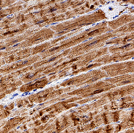
Supportive validation
- Submitted by
- R&D Systems (provider)
- Main image

- Experimental details
- Myoglobin in Human Heart. Myoglobin was detected in immersion fixed paraffin-embedded sections of human heart using Rabbit Anti-Human Myoglobin Monoclonal Antibody (Catalog # MAB97203) at 3 µg/mL for 1 hour at room temperature followed by incubation with the Anti-Rabbit IgG VisUCyte™ HRP Polymer Antibody (Catalog # VC003). Tissue was stained using DAB (brown) and counterstained with hematoxylin (blue). Specific staining was localized to cytoplasm in cardiomyocytes. View our protocol for IHC Staining with VisUCyte HRP Polymer Detection Reagents.