 Explore
 Validate
 Learn
Learn Immunocytochemistry
Antibody data
- Antibody Data
- Antigen structure
- References [10]
- Comments [0]
- Validations
- Immunocytochemistry [1]
- Immunohistochemistry [1]
Submit
Validation data
Reference
Comment
Report error
- Product number
- HPA012867 - Provider product page

- Provider
- Atlas Antibodies
- Proper citation
- Atlas Antibodies Cat#HPA012867, RRID:AB_1856710
- Product name
- Anti-HTR2B
- Antibody type
- Polyclonal
- Description
- Polyclonal Antibody against Human HTR2B, Gene description: 5-hydroxytryptamine (serotonin) receptor 2B, G protein-coupled, Alternative Gene Names: 5-HT(2B), 5-HT2B, Validated applications: ICC, IHC, Uniprot ID: P41595, Storage: Store at +4°C for short term storage. Long time storage is recommended at -20°C.
- Reactivity
- Human
- Host
- Rabbit
- Conjugate
- Unconjugated
- Isotype
- IgG
- Vial size
- 100 µl
- Concentration
- 0.1 mg/ml
- Storage
- Store at +4°C for short term storage. Long time storage is recommended at -20°C.
- Handling
- The antibody solution should be gently mixed before use.
Submitted references Genomic landscape and expression profile of consensus molecular subtype four of colorectal cancer
Molecular and clinicopathological differences between depressed and protruded T2 colorectal cancer
Fibroblast activation protein identifies Consensus Molecular Subtype 4 in colorectal cancer and allows its detection by 68Ga-FAPI-PET imaging
Stromal SOX2 Upregulation Promotes Tumorigenesis through the Generation of a SFRP1/2-Expressing Cancer-Associated Fibroblast Population
PD-L1 as a Prognostic Factor in Early-Stage Colon Carcinoma within the Immunohistochemical Molecular Subtype Classification
Immunohistochemistry-Based Consensus Molecular Subtypes as a Prognostic and Predictive Biomarker for Adjuvant Chemotherapy in Patients with Stage II Colorectal Cancer
Early-stage serrated adenocarcinomas are divided into several molecularly distinct subtypes
Collagen-rich stroma in aggressive colon tumors induces mesenchymal gene expression and tumor cell invasion
Human Polyomavirus Receptor Distribution in Brain Parenchyma Contrasts with Receptor Distribution in Kidney and Choroid Plexus
Poor-prognosis colon cancer is defined by a molecularly distinct subtype and develops from serrated precursor lesions
Lu Y, Gu D, Zhao C, Sun Y, Li W, He L, Wang X, Kou Z, Su J, Guo F
Frontiers in Immunology 2023;14
Frontiers in Immunology 2023;14
Molecular and clinicopathological differences between depressed and protruded T2 colorectal cancer
Cheah P, Mochizuki K, Kudo S, Kato K, Kudo K, Ogawa Y, Kouyama Y, Takashina Y, Ichimasa K, Tobo T, Toshima T, Hisamatsu Y, Yonemura Y, Masuda T, Miyachi H, Ishida F, Nemoto T, Mimori K
PLOS ONE 2022;17(10):e0273566
PLOS ONE 2022;17(10):e0273566
Fibroblast activation protein identifies Consensus Molecular Subtype 4 in colorectal cancer and allows its detection by 68Ga-FAPI-PET imaging
Strating E, Wassenaar E, Verhagen M, Rauwerdink P, van Schelven S, de Hingh I, Rinkes I, Boerma D, Witkamp A, Lacle M, Fodde R, Volckmann R, Koster J, Stedingk K, Giesel F, de Roos R, Poot A, Bol G, Lam M, Elias S, Kranenburg O
British Journal of Cancer 2022;127(1):145-155
British Journal of Cancer 2022;127(1):145-155
Stromal SOX2 Upregulation Promotes Tumorigenesis through the Generation of a SFRP1/2-Expressing Cancer-Associated Fibroblast Population
Kasashima H, Duran A, Martinez-Ordoñez A, Nakanishi Y, Kinoshita H, Linares J, Reina-Campos M, Kudo Y, L’Hermitte A, Yashiro M, Ohira M, Bao F, Tauriello D, Batlle E, Diaz-Meco M, Moscat J
Developmental Cell 2021;56(1):95-110.e10
Developmental Cell 2021;56(1):95-110.e10
PD-L1 as a Prognostic Factor in Early-Stage Colon Carcinoma within the Immunohistochemical Molecular Subtype Classification
Azcue P, Encío I, Guerrero Setas D, Suarez Alecha J, Galbete A, Mercado M, Vera R, Gomez-Dorronsoro M
Cancers 2021;13(8):1943
Cancers 2021;13(8):1943
Immunohistochemistry-Based Consensus Molecular Subtypes as a Prognostic and Predictive Biomarker for Adjuvant Chemotherapy in Patients with Stage II Colorectal Cancer
Li Y, Yao Q, Zhang L, Mo S, Cai S, Huang D, Peng J
The Oncologist 2020;25(12):e1968-e1979
The Oncologist 2020;25(12):e1968-e1979
Early-stage serrated adenocarcinomas are divided into several molecularly distinct subtypes
Katoh M, Hirano D, Urabe Y, Tanaka S, Nakamura K, Ninomiya Y, Yuge R, Hayashi R, Oka S, Kitadai Y, Shimamoto F, Arihiro K, Chayama K
PLOS ONE 2019;14(2):e0211477
PLOS ONE 2019;14(2):e0211477
Collagen-rich stroma in aggressive colon tumors induces mesenchymal gene expression and tumor cell invasion
Vellinga T, den Uil S, Rinkes I, Marvin D, Ponsioen B, Alvarez-Varela A, Fatrai S, Scheele C, Zwijnenburg D, Snippert H, Vermeulen L, Medema J, Stockmann H, Koster J, Fijneman R, de Rooij J, Kranenburg O
Oncogene 2016;35(40):5263-5271
Oncogene 2016;35(40):5263-5271
Human Polyomavirus Receptor Distribution in Brain Parenchyma Contrasts with Receptor Distribution in Kidney and Choroid Plexus
Haley S, O'Hara B, Nelson C, Brittingham F, Henriksen K, Stopa E, Atwood W
The American Journal of Pathology 2015;185(8):2246-2258
The American Journal of Pathology 2015;185(8):2246-2258
Poor-prognosis colon cancer is defined by a molecularly distinct subtype and develops from serrated precursor lesions
De Sousa E Melo F, Wang X, Jansen M, Fessler E, Trinh A, de Rooij L, de Jong J, de Boer O, van Leersum R, Bijlsma M, Rodermond H, van der Heijden M, van Noesel C, Tuynman J, Dekker E, Markowetz F, Medema J, Vermeulen L
Nature Medicine 2013;19(5):614-618
Nature Medicine 2013;19(5):614-618
No comments: Submit comment
Supportive validation
- Submitted by
- Atlas Antibodies (provider)
- Main image
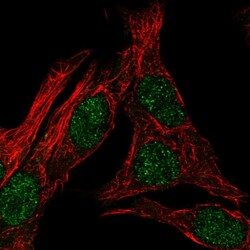
- Experimental details
- Immunofluorescent staining of human cell line SK-MEL-30 shows localization to nucleoplasm.
- Sample type
- Human

Supportive validation
- Submitted by
- Atlas Antibodies (provider)
- Enhanced method
- Orthogonal validation
- Main image
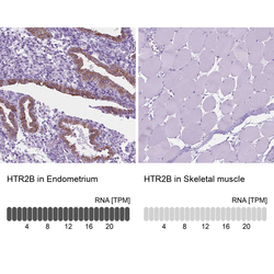
- Experimental details
- Immunohistochemistry analysis in human endometrium and skeletal muscle tissues using HPA012867 antibody. Corresponding HTR2B RNA-seq data are presented for the same tissues.
- Sample type
- Human
- Protocol
- Protocol
