Explore
 Validate
 Learn
LearnMAB590-100
antibody from R&D Systems
Targeting: CCR6
BN-1, CD196, CKR-L3, CMKBR6, DCR2, DRY-6, GPR-CY4, GPR29, STRL22
 Immunohistochemistry
Antibody data
- Antibody Data
- Antigen structure
- References [7]
- Comments [0]
- Validations
- Immunohistochemistry [2]
- Flow cytometry [1]
Submit
Validation data
Reference
Comment
Report error
- Product number
- MAB590-100 - Provider product page

- Provider
- R&D Systems
- Product name
- Mouse CCR6 Antibody
- Antibody type
- Monoclonal
- Description
- Protein A or G purified from hybridoma culture supernatant. Detects mouse CCR6-transfected rat RBL cells but not the parent cell line or cells transfected with mouse CCR5.
- Reactivity
- Mouse
- Host
- Rat
- Conjugate
- Unconjugated
- Antigen sequence
O54689- Isotype
- IgG
- Antibody clone number
- 140706
- Vial size
- 100 ug
- Concentration
- LYOPH
- Storage
- Use a manual defrost freezer and avoid repeated freeze-thaw cycles. 12 months from date of receipt, -20 to -70 °C as supplied. 1 month, 2 to 8 °C under sterile conditions after reconstitution. 6 months, -20 to -70 °C under sterile conditions after reconstitution.
Submitted references IL-1β and IL-23 Promote Extrathymic Commitment of CD27+CD122- γδ T Cells to γδT17 Cells.
IL-10+ Innate-like B Cells Are Part of the Skin Immune System and Require α4β1 Integrin To Migrate between the Peritoneum and Inflamed Skin.
CCR6 is a prognostic marker for overall survival in patients with colorectal cancer, and its overexpression enhances metastasis in vivo.
SerpinB1 regulates homeostatic expansion of IL-17+ γδ and CD4+ Th17 cells.
Colonic patch and colonic SILT development are independent and differentially regulated events.
CCR6 is required for epidermal trafficking of γδ-T cells in an IL-23-induced model of psoriasiform dermatitis.
Human beta-defensin 2 and 3 and their mouse orthologs induce chemotaxis through interaction with CCR2.
Muschaweckh A, Petermann F, Korn T
Journal of immunology (Baltimore, Md. : 1950) 2017 Oct 15;199(8):2668-2679
Journal of immunology (Baltimore, Md. : 1950) 2017 Oct 15;199(8):2668-2679
IL-10+ Innate-like B Cells Are Part of the Skin Immune System and Require α4β1 Integrin To Migrate between the Peritoneum and Inflamed Skin.
Geherin SA, Gómez D, Glabman RA, Ruthel G, Hamann A, Debes GF
Journal of immunology (Baltimore, Md. : 1950) 2016 Mar 15;196(6):2514-2525
Journal of immunology (Baltimore, Md. : 1950) 2016 Mar 15;196(6):2514-2525
CCR6 is a prognostic marker for overall survival in patients with colorectal cancer, and its overexpression enhances metastasis in vivo.
Liu J, Ke F, Xu Z, Liu Z, Zhang L, Yan S, Wang Z, Wang H, Wang H
PloS one 2014;9(6):e101137
PloS one 2014;9(6):e101137
SerpinB1 regulates homeostatic expansion of IL-17+ γδ and CD4+ Th17 cells.
Zhao P, Hou L, Farley K, Sundrud MS, Remold-O'Donnell E
Journal of leukocyte biology 2014 Mar;95(3):521-30
Journal of leukocyte biology 2014 Mar;95(3):521-30
Colonic patch and colonic SILT development are independent and differentially regulated events.
Baptista AP, Olivier BJ, Goverse G, Greuter M, Knippenberg M, Kusser K, Domingues RG, Veiga-Fernandes H, Luster AD, Lugering A, Randall TD, Cupedo T, Mebius RE
Mucosal immunology 2013 May;6(3):511-21
Mucosal immunology 2013 May;6(3):511-21
CCR6 is required for epidermal trafficking of γδ-T cells in an IL-23-induced model of psoriasiform dermatitis.
Mabuchi T, Singh TP, Takekoshi T, Jia GF, Wu X, Kao MC, Weiss I, Farber JM, Hwang ST
The Journal of investigative dermatology 2013 Jan;133(1):164-71
The Journal of investigative dermatology 2013 Jan;133(1):164-71
Human beta-defensin 2 and 3 and their mouse orthologs induce chemotaxis through interaction with CCR2.
Röhrl J, Yang D, Oppenheim JJ, Hehlgans T
Journal of immunology (Baltimore, Md. : 1950) 2010 Jun 15;184(12):6688-94
Journal of immunology (Baltimore, Md. : 1950) 2010 Jun 15;184(12):6688-94
No comments: Submit comment
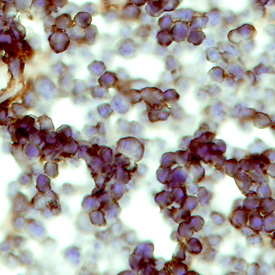
Supportive validation
- Submitted by
- R&D Systems (provider)
- Main image

- Experimental details
- CCR6 in Mouse Thymus. CCR6 was detected in perfusion fixed frozen sections of mouse thymus using Mouse CCR6 Monoclonal Antibody (Catalog # MAB590) at 8 µg/mL overnight at 4 °C. Tissue was stained using the Anti-Rat HRP-DAB Cell & Tissue Staining Kit (brown; Catalog # CTS017) and counterstained with hematoxylin (blue). Specific labeling was localized to the plasma membrane of lymphocytes. View our protocol for Chromogenic IHC Staining of Frozen Tissue Sections.
- Submitted by
- R&D Systems (provider)
- Main image
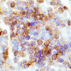
- Experimental details
- CCR6 in Mouse Spleen. CCR6 was detected in immersion fixed frozen sections of mouse spleen using Rat Anti-Mouse CCR6 Monoclonal Antibody (Catalog # MAB590) at 0.3 µg/mL overnight at 4 °C. Tissue was stained using the Anti-Rat HRP-DAB Cell & Tissue Staining Kit (brown; CTS017) and counterstained with hematoxylin (blue). Specific staining was localized to plasma membranes in lymphocytes. View our protocol for Chromogenic IHC Staining of Frozen Tissue Sections.
Supportive validation
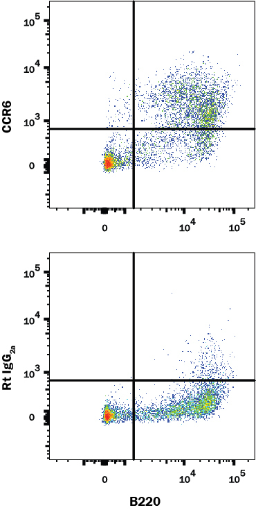
- Submitted by
- R&D Systems (provider)
- Main image
- Experimental details
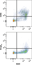
- Detection of CCR6 in Mouse Splenocytes by Flow Cytometry. Mouse splenocytes were stained with (A) Rat Anti-Mouse CCR6 Monoclonal Antibody (Catalog # MAB590) or (B) Rat IgG2A control antibody (Catalog # MAB006) followed by Goat anti-Rat IgG APC-conjugated Secondary Antibody (Catalog # F0113) and Rat Anti-Mouse B220/CD45R Fluorescein-conjugated Monoclonal Antibody (Catalog # FAB1217F). View our protocol for Staining Membrane-associated Proteins.