 Explore
 Validate
 Learn
Learn25-1969-41
antibody from Invitrogen Antibodies
Targeting: CCR6
BN-1, CD196, CKR-L3, CMKBR6, DCR2, DRY-6, GPR-CY4, GPR29, STRL22
 Flow cytometry
Antibody data
- Antibody Data
- Antigen structure
- References [7]
- Comments [0]
- Validations
- Flow cytometry [1]
Submit
Validation data
Reference
Comment
Report error
- Product number
- 25-1969-41 - Provider product page

- Provider
- Invitrogen Antibodies
- Product name
- Anti-CD196 (CCR6) Monoclonal Antibody (R6H1), PE-Cyanine7, eBioscience™
- Antibody type
- Monoclonal
- Antigen
- Other
- Description
- Description: This R6H1 monoclonal antibody reacts with CD196 (also known as CCR6), a seven transmembrane G protein-coupled receptor expressed on T, B, dendritic, natural killer, and Langerhans cells. This CC chemokine receptor uniquely binds MIP-3a/CCL20, a chemoattractant for dendritic cells, effector/memory T cells, and B cells. CD196 is also involved in host defense and inflammation at epithelial sites. Furthermore, this receptor has been implicated in Th17 differentiation and CD4+FoxP3+ regulatory T cell development. Applications Reported: This R6H1 antibody has been reported for use in flow cytometric analysis. Applications Tested: This R6H1 antibody has been pre-titrated and tested By flow cytometric analysis on normal human peripheral blood cells. This can be used at 5 µL (0.125 µg) per test. A test is defined as the amount (µg) of antibody that will stain a cell sample in a final volume of 100 µL. Cell number should be determined empirically but can range from 10^5 to 10^8 cells/test. Light sensitivity: This tandem dye is sensitive photo-induced oxidation. Please protect this vial and stained samples from light. Fixation: Samples can be stored in IC Fixation Buffer (cat. 00-8222) (100 µL cell sample + 100 µL IC Fixation Buffer) or 1-step Fix/Lyse Solution (cat. 00-5333) for up to 3 days in the dark at 4°C with minimal impact on brightness and FRET efficiency/compensation. Some generalizations regarding fluorophore performance after fixation can be made, but clone specific performance should be determined empirically. Excitation: 488-561 nm; Emission: 775 nm; Laser: Blue Laser, Green Laser, Yellow-Green Laser. Filtration: 0.2 µm post-manufacturing filtered.
- Reactivity
- Human
- Host
- Mouse
- Isotype
- IgG
- Antibody clone number
- R6H1
- Vial size
- 25 Tests
- Concentration
- 5 µL/Test
- Storage
- 4° C, store in dark, DO NOT FREEZE!
Submitted references Survival, Retention, and Selective Proliferation of Lymphocytes Is Mediated by Gingival Fibroblasts.
Differential adipokine receptor expression on circulating leukocyte subsets in lean and obese children.
Fluctuations in Blood Marginal Zone B-Cell Frequencies May Reflect Migratory Patterns Associated with HIV-1 Disease Progression Status.
Identification and characterization of latency-associated peptide-expressing γδ T cells.
CCL20 Secretion from the Nucleus Pulposus Improves the Recruitment of CCR6-Expressing Th17 Cells to Degenerated IVD Tissues.
IL-7 licenses activation of human liver intrasinusoidal mucosal-associated invariant T cells.
Down-regulation of the beta-chemokine receptor CCR6 in dendritic cells mediated by TNF-alpha and IL-4.
Moonen CGJ, Alders ST, Bontkes HJ, Schoenmaker T, Nicu EA, Loos BG, de Vries TJ
Frontiers in immunology 2018;9:1725
Frontiers in immunology 2018;9:1725
Differential adipokine receptor expression on circulating leukocyte subsets in lean and obese children.
Keustermans G, van der Heijden LB, Boer B, Scholman R, Nuboer R, Pasterkamp G, Prakken B, de Jager W, Kalkhoven E, Janse AJ, Schipper HS
PloS one 2017;12(10):e0187068
PloS one 2017;12(10):e0187068
Fluctuations in Blood Marginal Zone B-Cell Frequencies May Reflect Migratory Patterns Associated with HIV-1 Disease Progression Status.
Gauvin J, Chagnon-Choquet J, Poudrier J, Roger M, Montreal Primary HIV Infection and Slow Progressor Cohorts.
PloS one 2016;11(5):e0155868
PloS one 2016;11(5):e0155868
Identification and characterization of latency-associated peptide-expressing γδ T cells.
Rezende RM, da Cunha AP, Kuhn C, Rubino S, M'Hamdi H, Gabriely G, Vandeventer T, Liu S, Cialic R, Pinheiro-Rosa N, Oliveira RP, Gaublomme JT, Obholzer N, Kozubek J, Pochet N, Faria AMC, Weiner HL
Nature communications 2015 Dec 8;6:8726
Nature communications 2015 Dec 8;6:8726
CCL20 Secretion from the Nucleus Pulposus Improves the Recruitment of CCR6-Expressing Th17 Cells to Degenerated IVD Tissues.
Zhang W, Nie L, Wang Y, Wang XP, Zhao H, Dongol S, Maharjan S, Cheng L
PloS one 2013;8(6):e66286
PloS one 2013;8(6):e66286
IL-7 licenses activation of human liver intrasinusoidal mucosal-associated invariant T cells.
Tang XZ, Jo J, Tan AT, Sandalova E, Chia A, Tan KC, Lee KH, Gehring AJ, De Libero G, Bertoletti A
Journal of immunology (Baltimore, Md. : 1950) 2013 Apr 1;190(7):3142-52
Journal of immunology (Baltimore, Md. : 1950) 2013 Apr 1;190(7):3142-52
Down-regulation of the beta-chemokine receptor CCR6 in dendritic cells mediated by TNF-alpha and IL-4.
Carramolino L, Kremer L, Goya I, Varona R, Buesa JM, Gutiérrez J, Zaballos A, Martínez-A C, Márquez G
Journal of leukocyte biology 1999 Nov;66(5):837-44
Journal of leukocyte biology 1999 Nov;66(5):837-44
No comments: Submit comment
Supportive validation
- Submitted by
- Invitrogen Antibodies (provider)
- Main image
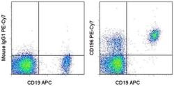
- Experimental details
- Staining of normal human peripheral blood cells with Anti-Human CD19 APC (Product # 17-0199-42) and Mouse IgG1 kappa Isotype Control PE-Cyanine7 (Product # 25-4714-80) (left) or Anti-Human CD196 (CCR6) PE-Cyanine7 (right). Cells in the lymphocyte gate were used for analysis.
