Explore
 Validate
 Learn
LearnPA5-29015
antibody from Invitrogen Antibodies
Targeting: CCR6
BN-1, CD196, CKR-L3, CMKBR6, DCR2, DRY-6, GPR-CY4, GPR29, STRL22
 Western blot
 Immunohistochemistry
Antibody data
- Antibody Data
- Antigen structure
- References [1]
- Comments [0]
- Validations
- Immunohistochemistry [2]
- Other assay [2]
Submit
Validation data
Reference
Comment
Report error
- Product number
- PA5-29015 - Provider product page

- Provider
- Invitrogen Antibodies
- Product name
- CCR6 Polyclonal Antibody
- Antibody type
- Polyclonal
- Antigen
- Synthetic peptide
- Description
- Recommended positive controls: 293T, A431, H1299, HeLaS3, HepG2, Molt-4, Raji. Store product as a concentrated solution. Centrifuge briefly prior to opening the vial.
- Reactivity
- Human
- Host
- Rabbit
- Isotype
- IgG
- Vial size
- 100 μL
- Concentration
- 1 mg/mL
- Storage
- Store at 4°C short term. For long term storage, store at -20°C, avoiding freeze/thaw cycles.
Submitted references Expression of CCL20 and Its Corresponding Receptor CCR6 Is Enhanced in Active Inflammatory Bowel Disease, and TLR3 Mediates CCL20 Expression in Colonic Epithelial Cells.
Skovdahl HK, Granlund Av, Østvik AE, Bruland T, Bakke I, Torp SH, Damås JK, Sandvik AK
PloS one 2015;10(11):e0141710
PloS one 2015;10(11):e0141710
No comments: Submit comment
Supportive validation
- Submitted by
- Invitrogen Antibodies (provider)
- Main image
- Experimental details
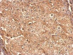
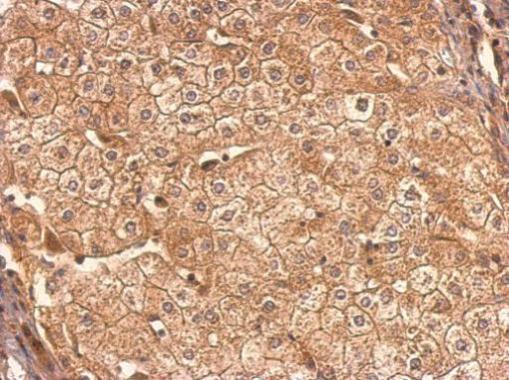
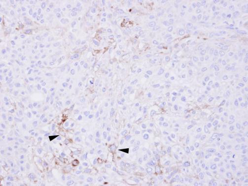
- CCR6 Polyclonal Antibody detects CCR6 protein at membrane on human hepatoma by immunohistochemical analysis. Sample: Paraffin-embedded hepatoma. CCR6 Polyclonal Antibody (Product # PA5-29015) dilution: 1:1,000. Antigen Retrieval: EDTA based buffer, pH 8.0, 15 min.
- Submitted by
- Invitrogen Antibodies (provider)
- Main image
- Experimental details
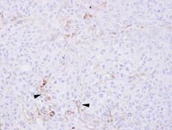
- Immunohistochemical analysis of paraffin-embedded DU-145 xenograft, using CCR6 (Product # PA5-29015) antibody at 1:100 dilution. Antigen Retrieval: EDTA based buffer, pH 8.0, 15 min.
Supportive validation
- Submitted by
- Invitrogen Antibodies (provider)
- Main image
- Experimental details
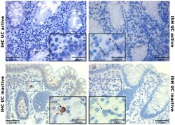
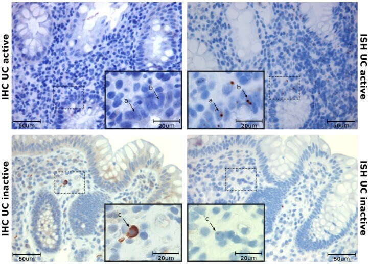
- Fig 5 CCR6 protein and mRNA in serial sections. Immunohistochemistry (IHC) and in situ hybridization (ISH) show CCR6 protein and mRNA in colonic biopsies from active UC and inactive CD. Serial sections from the same biopsy were used to compare the localization of mRNA and protein. Arrows show that there is no overlap of protein and mRNA in active disease (a, b), while a clear overlap is seen in inactive disease(c). Scale bars as indicated.
- Submitted by
- Invitrogen Antibodies (provider)
- Main image
- Experimental details
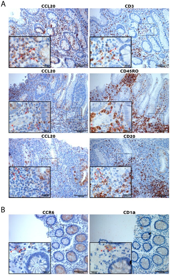
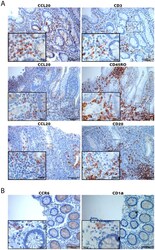
- Fig 6 Immune cell typing of CCL20 and CCR6 positive cells in serial sections. Immunohistochemistry on serial sections from the same biopsy was performed to localize CCR6 and CCL20 positivity to different immune cells. A: Sections from biopsies from active disease were used for CCL20 investigations. There is a partial overlap between CCL20+ cells and CD3+, CD45RO+ and CD20+ cells (arrows a, b, c, d). B: Sections from biopsies from inactive disease for CCR6. There is a partial overlap between CCR6+ cells and CD1alpha+ cells (arrows a, b). Scale bars as indicated.